| Клитор | |
|---|---|
| лат. clitoris | |
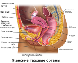 Женские тазовые органы в продольном разрезе |
|
 Строение клитора |
|
| Система | Репродуктивная система человека > Женская репродуктивная система > Наружные половые органы женщины |
| Кровоснабжение | дорсальная и глубокая артерии клитора[1] |
| Венозный отток | поверхностные дорсальные вены клитора, глубокая дорсальная вена клитора[2] |
| Иннервация | ветви дорсальных и пещеристых нервов клитора[3] |
| Прекурсор | Генитальный бугорок |
| Каталоги | |
|
|
Кли́тор (лат. clitoris от лат. clitorido — «щекочу»; также встречается термин «похотник»[4]; старослав. сикель (также секиль, секель)[5][6][7][8]) — непарный половой орган у самок млекопитающих, имеющий функцию одной из их главных эрогенных зон.
Входит в число женских наружных половых органов — наружных органов женской репродуктивной (мочеполовой) системы наряду с большими (наружными) и малыми половыми губами, находящимися внутри больших, с которыми имеет общие элементы кровотока и иннервации, однако в отличие от половых губ не имеет как целое покровной функции, а является преимущественно органом полового чувства[9].
Расположен позади и ниже передней спайки больших половых губ. Внутренняя структура клитора имеет форму перевёрнутой латинской буквы Y, немного сдавлен с боков. Состоит из головки (лат. glans clitoridis), тела (лат. corpus clitoridis) и двух ножек (лат. crura clitoridis), из числа которых обычно снаружи наблюдается только головка и иногда часть тела клитора, прикрытые кожной складкой — крайней плотью клитора.
При половом возбуждении образующие клитор два продольных пещеристых тела наполняются кровью (эрекция клитора) подобно пещеристым телам пениса (мужского полового члена)[10]. Клитор, развиваясь из одних с ним эмбриональных тканей, является гомологом его пещеристых тел, но в норме значительно меньше его[11] и, в отличие от пениса, не содержит в своей структуре губчатое тело и проходящий в последнем мочеиспускательный канал, которые у женщин расположены позади клитора.
Клитор содержит множество сосудов и нервных окончаний и у женщин является одной из самых чувствительных эрогенных зон.
История медицинского изучения клитора
Терминология
История изучения клитора насчитывает множество «открытий» этой структуры исследователями разных стран и в разные века. Использовалась также разная терминология[12]. Гиппократ использовал термин columella (маленькая колонна). Авиценна называл клитор albatra или virga (стержень). Абулкасис, другой арабский медик, назвал его tentigo (напряжение). Реальдо Коломбо использовал термины amoris dulcedo (сладость любви), sedes libidinis (седалище похоти) и «муха Венеры»[13]. Средневековый схоластик Альберт Магнус подчёркивал гомологию между мужскими и женскими структурами, используя термин virga для обозначения как мужских, так и женских гениталий[14][15]. Древние римляне использовали неприличное слово landīca для обозначения клитора[16]. Слово было настолько грубым, что оно практически не встречается в рукописях, за исключением Priapeia 78, где используется выражение misella landica («бедный клитор»)[17]. Слово, однако, присутствует в граффити: peto [la]ndicam fvlviae («Я ищу клитор Фульвии»), нанесённом на свинцовый метательный снаряд, найденный в Перудже[18].
Ренье де Грааф подчёркивал, что следует отличать нимфу от клитора, поэтому он предложил называть структуру только клитором[19]. Начиная с XVII века это название стало общепринятым, нимфой же сначала называли вульву, а позже малые половые губы[20]. Греческое слово κλειτορίς, возможно происходит от «щекотать»[13], но оно также может обозначать «маленькую горку»; то есть древние авторы могли воспользоваться игрой слов[21]. Лингвист Марсель Коэн посвятил главу в книге исследованию происхождения слова «клитор», но не пришёл к определённым выводам[22].
Открытия и повторные открытия
Из-за его весьма малых размеров и скрытого расположения большей части его структуры в окружающих складках жировых и кожных покровов клитор часто малодоступен непосредственному наблюдению, поэтому можно вести речь об истории его открытия, которому не способствовало и отсутствие у него собственно репродуктивной функции.
Открытие клитора часто связывают с именем итальянского анатома XVI века Реальдо Коломбо[23]. В 1559 году он издал книгу «De anatomica», в которой описал «женское место удовольствия при сношении» и назвал себя первооткрывателем этого органа. Коломбо писал:
Так как никто не описал эти отростки и их функционирование, и если позволительно давать название органам, обнаруженным мной, то следует назвать его любовью или сладостью Венеры…
Коломбо также упомянул клитор в разделе о редких анатомических образованиях — он описал эфиопскую женщину, клитор у которой был длиной и толщиной с мизинец, а вагинальное отверстие было очень узким[24].
Андреас Везалий — друг и учитель Коломбо, отношения с которым затем были испорчены — отверг это открытие. Везалий полагал, что женские гениталии представляют собой симметричное отображение мужских. Следуя этой теории, пенису ставилось в соответствие влагалище, а для клитора не было соответствующего мужского органа. Возражая против идей Коломбо, Везалий писал:
Не имеет смысла обвинять других в некомпетентности на основании игры природы, которые вы наблюдали у некоторых женщин, и вы не сможете определить эту новую и ненужную часть у здоровых женщин. Я полагаю, что такая структура имеется у гермафродитов, у которых ярко выражены гениталии, как описывает Павел Эгинский, но я ни разу не видел ни у одной женщины пениса (который Авиценна назвал албарата и греки называли увеличенной нимфой и классифицировали как заболевание) или даже рудимента мельчайшего фаллоса[25][26].
Первенство Коломбо в открытии клитора было оспорено его преемником в Падуе, Габриеле Фаллопием, который назвал первооткрывателем самого себя. В своём труде «Observations anatomicae», написанном в 1550-е годы и опубликованном в 1561 году, он отметил, что эта часть женской анатомии настолько труднообнаружима, что он оказался первым, кто открыл её; прочие же сообщали об этом органе либо с его слов, либо со слов его студентов[27].
Каспар Бартолин, анатом XVII века, отклонил оба заявления, утверждая, что клитор был широко известен медицинской науке ещё со II века[28].
До Коломбо и Фаллопио клитор описывали древнегреческие, персидские и арабские медики и хирурги[29][30], но его функция трактовалась ошибочно. Французский анатом Шарль Эстьен в труде 1545 года «De Dissectione Partium Corporis Humani» приписал клитору (membre honteux, срамному члену) роль в мочеиспускании[31]. Возможно, Коломбо первый описал сексуальную функцию клитора, но даже это оспаривается.

Conciliator differentiarum philosophorum et precipue medicorum
Итальянский философ и профессор медицины Пьетро д’Абано в книге «Conciliator differentiarum philosophorum et medicorum» (1476) писал, что трение верхнего орифиса около лобка вызывает возбуждение у женщин, хотя и не изучил детальную анатомию клитора[30].
Инки, XV—XVI века
Хирургические операции над клитором осуществлялись в Империи Инков. В столице инков — городе Куско — существовал храм Амаруканча (в XV—XVI веках; позднее — в последней трети XVI века — в нём разместились иезуиты), назначением которого, согласно записям итальянского священника, иезуита XVII века Хуана Анелло Оливы, было
поклонение идолу в виде дракона-змея, пожирающего скорпиона.
С этим местом связана одна интересная деталь сакральных верований и философии инков, приводимая тем же автором:
Дракон был жизненной силой Создателя; и как мы, католики, почитаем женский облик Божества Святой Девы Марии, подобным же образом вместилище и жало скорпиона у [Инков] символизировали женский клитор, почитая в нём мужское начало в женщине
Поэтому у инков бытовал обычай обрезать клитор у девочек, что католическими священниками расценивалось как варварство[32].
Анатомия

Строение клитора и смежных структур. Наружные и внутренние части клитора находятся сверху и выделены темно-розовым цветом: glans — округлая головка клитора, венчающая его тело и выступающая наружу вперед из-под его крайней плоти; corpus cavernosum (лат.) — одно из двух пещеристых тел, образующих тело клитора; crus (лат.) — одна из двух ножек клитора. Внизу рисунка вход в более широкий трубчатый орган — влагалище (англ. vaginal opening), а над ним более узкий — в уретру (англ. urethral opening); по бокам от них луковицы преддверия влагалища (англ. bulbs of vestibule). Контурами обозначены большие и малые половые губы, переходящие в верхней части в крайнюю плоть клитора

Клитор, согласно исследованиям австралийского уролога, доктора Хелен О’Коннелл, состоит из двух пещеристых тел клитора (лат. corpus cavernosum clitoridis), головки клитора (лат. glans clitoridis), ножек клитора (лат. crus clitoridis) и двух луковиц преддверия влагалища (иначе клиторальные луковицы) (лат. bulbus vestibuli vaginae)[33]. Волокнистые мембраны, окружающие пещеристые половинки тела клитора, сходятся над срединными поверхностями, образуя перегородку, к которой прикрепляются эластичные и гладкомышечные волокна. Пещеристое тело клитора разделяется над уретрой на две ножки клитора, которые огибают с двух сторон уретру и вагину, и оканчиваются двумя луковицами образуя клитороуретровагинальный комплекс. Тело клитора связано с седалищно-лобковой ветвью (лат. ramus ischiopubicus) корнем, в то время как две небольшие седалищно-пещеристые мышцы (лат. musculus ischiocavernosus) прикрепляются к ножкам клитора во внутренней части головки и пещеристых тел, образуя комплекс нервных окончаний. Кровоснабжение клитора обеспечивают ветви внутренней срамной артерии (лат. arteria pudenda interna). Приток артериальной крови и венозный отток одинаковы с мужским половым членом[10].
Можно выделить три основные зоны видимой части клитора: головка, уздечка и крайняя плоть клитора (клиторальный капюшон).
С точки зрения анатомии клитор равнозначен мужскому половому члену за исключением отсутствия в структуре клитора мочеиспускательного канала и губчатого тела.
Головка клитора
Головка клитора (лат. glans clitoridis) — одна из самых чувствительных частей женского тела, она насыщена кровеносными сосудами и нервными окончаниями (тельцами Пачини, Мейснера, Краузе, Догеля). У некоторых женщин головка настолько чувствительна, что её прямая стимуляция (при мастурбации или куннилингусе) может вызвать неприятные ощущения. Головка прикрыта кожной складкой (крайней плотью или так называемым клиторальным капюшоном (англ. clitoral hood)) . В спокойном состоянии головка либо не видна вовсе, либо видна лишь небольшая её часть. При половом возбуждении происходит эрекция клитора, головка выступает вперёд.
Головка клитора развивается из тех же тканей[11], что и головка мужского полового члена, являясь гомологичной ей генетически структурой, хотя головка пениса представляет собой губчатую ткань, продолжающую губчатое тело пениса, а головка клитора является оконечностью его пещеристых тел. Головка клитора тоже богата чувствительными нервными окончаниями и способна к кровенаполнению при эрекции, но значительно меньше её и, в отличие от пениса, не имеет в своей толще губчатого тела и проходящей в нём трубчатой полости уретры, а на своей поверхности соответственно не несёт наружного отверстия, в которое у мужчин открывается мочеиспускательный канал, служащий для выведения из организма мужчины как мочи (мочеиспускание), так и семенной жидкости (эякуляция), а у женщин для мочеиспускания.
Головку клитора так же, как и пениса, защищает от внешних воздействий относительно подвижная кожная складка — крайняя плоть, внутренний листок которой богат вырабатывающими густой секрет, защищающий головку от сухости — смегму, скопления которой при несоблюдении личной гигиены могут приводить к раздражению и воспалению.
Крайняя плоть клитора

Пирсинг клиторального капюшона
Крайняя плоть клитора (лат. preputium clitoridis) — кожная складка, прикрывающая наружную часть тела клитора и часто его головку для их защиты от внешних повреждений; от сухости головку защищают своими выделениями — смегмой, расположенные на внутреннем листке крайней плоти сальные железы. Она по своему происхождению и покровной функции аналогична крайней плоти мужского полового члена, прикрывающей в невозбужденном состоянии его головку. В отличие от имеющего значительно большую длину пениса, у которого крайней плотью называют лишь дальнюю от тела мужчины часть его кожного покрова, крайней плотью клитора именуют весь небольшой кожный покров его наружной части.
Крайняя плоть, как и сама наружная часть клитора, имеет небольшие размеры, но обычно видна при наружном осмотре, хотя у некоторых женщин, имеющих пухлые большие половые губы, клитор практически не виден.
Крайняя плоть нередко может быть несколько отведена назад, в том числе при половом возбуждении, эрекции клитора, обнажая обладающую большим числом чувствительных нервных окончаний головку клитора, являющуюся одной из главных эрогенных зон. Отведение крайней плоти назад и обнажение головки бывает необходимо для удаления скоплений смегмы, которые могут при несоблюдении норм личной гигиены вызвать дискомфорт и воспаление.
Крайняя плоть клитора представляет собой самое популярное место женского интимного пирсинга. Чаще всего, когда сообщают о проколотом клиторе, имеется в виду именно горизонтальный прокол крайней плоти клитора, украшенный колечком, барбеллом[34], микробананчиком и проч. Прокол же клитора является одним из самых сложных и возможен только в случае биологической совместимости с данным видом пирсинга (маленький, плохо различимый клитор прокалывать нельзя).
Для увеличения чувствительности клитора может проводиться его хирургическое обнажение, предусматривающее удаление его крайней плоти, аналогичное обрезанию крайней плоти у мужчин.
Уздечка клитора
Продольная кожная складка, соединяющая передние концы малых половых губ с нижней поверхностью головки клитора м крайней плотью, называется уздечкой клитора (лат. frenulum clitoridis). Она натягивается при его половом возбуждении, как и аналогичная уздечка мужского полового члена, расположенная под его головкой и соединённая с его крайней плотью, и, пригибая головку, способствует её максимальной стимуляции.
Клитор во время полового акта
Клитор до (слева) и во время (справа) полового возбуждения
У большинства женщин клитор является главной эрогенной зоной. По этой причине именно клитор является основным источником приятных ощущений, которые женщина испытывает во время полового акта. В то же время при вагинальном коитусе член мужчины напрямую на головку клитора не воздействует, так движения мужского органа происходят во влагалище, воздействие производится на внутреннюю часть клитора опосредованно.
Стимуляция головки клитора во время полового акта происходит через прилегающие к нему части женских интимных органов, например, через натяжение и подёргивание малых половых губ. Обычно этого достаточно для нарастания возбуждения и достижения оргазма, но в ряде случаев женщины вынуждены прибегать к дополнительной стимуляции.
Как правило, клитор возбуждается не сразу. Это заметно по отсутствию секреторной жидкости, выделяющейся из половых органов женщины. В обычном случае сексуальное возбуждение сопровождается обильным выделением секреторной жидкости.
Незадолго до оргазма клитор уменьшается в размерах[источник не указан 3484 дня]. Имеется спорная точка зрения, согласно которой это частично защищает его чувствительную часть от дальнейших стимулов[35]. Спустя 5—10 секунд после оргазма клитор возвращается к своим нормальным размерам.
В момент оргазма ритмичные сокращения мускулов происходят во внешней трети половых органов и в матке. Они происходят первоначально приблизительно каждые 0,8 секунды, становясь менее интенсивными и более беспорядочно раздельными, поскольку оргазм продолжается. Оргазм может иметь разное число мышечных сокращений, в зависимости от интенсивности.
Немедленно после оргазма клитор может быть настолько чувствительным, что любая его стимуляция может вызывать дискомфорт и/или боль.
У некоторых женщин при половом возбуждении головка клитора может увеличиваться примерно вдвое, у других практически не меняет размера. В отличие от быстрой эрекции мужского полового члена, реакция клитора на половой стимул проявляется лишь спустя 20—30 секунд после начала воздействия.
При продолжительном интенсивном возбуждении головка может быть почти полностью скрыта в складках набухших малых половых губ.
Роль в достижении оргазма
|
|
Этот раздел статьи ещё не написан. Здесь может располагаться отдельный раздел. Помогите Википедии, написав его. (14 июля 2021) |
Размеры клитора

Соотношение размеров взрослых невозбужденного мужского полового члена (слева) и клитора (справа). Обведены рамкой. Преобладающая по размеру часть клитора является внутренней.
Размеры клитора задаются генетически и уровнем половых гормонов. Он развивается из тех же эмбриональных тканей, что и мужской половой член, но половая дифференциация благодаря воздействию половых гормонов в норме ведёт к тому, что размеры клитора и пениса занимают противоположное положение на шкале дифференциации полов Прадера, где пенис максимален, а клитор минимален. Под действием мужских гормонов — андрогенов — при нормальной работе их рецепторов пенис значительно вырастает в размерах, а тело и головка клитора без влияния андрогенов остаются маленькими, тогда как при генетических и гормональных отклонениях различие между пенисом и клитором в размерах, а иногда и структуре снижается, и при повышенной выработке мужских гормонов клитор может в широких пределах увеличиваться (гипертрофия клитора), что затрудняет определение пола у новорожденных[11]. Различие в размерах этих органов в норме определяется отличием ряда функций: мужской половой член призван доносить генетический материал мужчины во внутренние половые органы женщины, тогда как у клитора аналогичная функция отсутствует и при обычных размерах для проникновения он не предназначен. Необычно большие размеры клитора называют клиторомегалией, тогда как аномалии пениса в отношении его величины связаны, наоборот, с аномально малыми его размерами (микропенией).
Тео Ланг упоминает один зарегистрированный случай, когда у женщины головка клитора была 5 см в длину и достигала 7,5 см, «когда клитор находился в состоянии полной эрекции»[источник не указан 2010 дней]. Ральф Померой отмечал, что у белых женщин размеры головки клитора более 2,5 см в длину весьма редки, но встречаются у двух-трёх процентов чернокожих — «размеры 7,5 см и более выявляются примерно у каждой из 300 или 400 чернокожих женщин»[источник не указан 2722 дня]. Другой автор замечает, что Паран-Дюшателе встречал женщину, головка клитора которой имела в длину 8 см[источник не указан 2010 дней]. Швейцарский биолог XVIII века Альбрехт фон Галлер утверждал, что встречал женщину с огромным клитором не менее 18 см в длину[источник не указан 2010 дней].
Размеры клитора и его видимой части (головки) индивидуальны: общая длина головки от 4—5 мм до 1 см, диаметр от 2 до 20 мм. Полная длина клитора, включая его внутреннюю часть, обычно составляет от 8 до 20 см[36].
Вопреки установившемуся в некоторых кругах мнению, размеры клитора никак не связаны со степенью сексуального возбуждения, которое может испытывать женщина.
Нет никакой корреляции между размером клитора и возрастом, в том числе и с менопаузой и периодом после неё. Среди рожавших женщин, как правило, измерения дают несколько большие средние значения.
Гипертрофия клитора
Гипертрофия клитора наблюдается в случаях эмбриональных изменений андрогенов и сопровождается гиперсексуальностью. Причина гипертрофии — обычно это результат врождённых дефицитов надпочечных ферментов синтеза кортизола; более редко, это вызвано прогестационными агентами (утеро). Крайне редко, случается гипертрофия клитора, вызванная нейрофибромами клиториального тела, включая случай ограниченного нейрофибромного проникновения крайней плоти.
Лечение зависит от степени изменений, степени гипертрофии клитора и, в случае необходимости, на каком уровне влагалище поступает в мочеполовой синус.
(См. также Гермафродитизм)
Хирургия клитора
Клитор и другие женские наружные половые органы могут подвергаться хирургическим вмешательствам как в медицинских, так и в эстетических или в ритуальных целях. Тогда как медицинские вмешательства могут производиться в любой стране мира, ритуальные характерны для некоторых регионов Африки[37] и Азии. Чаще всего ритуальные вмешательства на женских наружных половых органах представляют собой различные виды женского обрезания, цель которого — уменьшение женской сексуальности для предотвращения промискуитета, внебрачных половых контактов. Для этого наружные половые органы лиц женского пола сшиваются с оставлением небольших выделительных отверстий (инфибуляция) или удаляются (клиторидэктомия). Женское обрезание широко критикуется как опасная для здоровья и дискриминационная практика.
Клиторидэктомия
Удаление клитора, а иногда и половых губ (частично или полностью). В качестве немедицинского ритуального вмешательства распространено в тех популяциях, где существует традиция женского обрезания и часто производится старшими женщинами рода или деревни. В качестве такового не вызвано медицинскими показаниями; часто выполняется в антисанитарных условиях, что чревато заражением крови с возможностью летального исхода, который может наступить и от болевого шока, так как анестезия обычно отсутствует. В результате этого ритуала подвергшаяся ему девочка или женщина лишается главной эрогенной зоны — клитора, а окружающие ткани наружных половых органов теряют естественную структуру и внешний вид и оказываются покрытыми болезненными и уродливыми шрамами, делающими половую жизнь женщины особенно болезненной и лишённой удовольствия.
В англоязычной литературе по этим вопросам помимо технического нейтрального обозначения этих ритуальных практик как female circumcision (женское обрезание) распространено их обозначение с негативной оценкой как female genital mutilation (FGM; нанесение женских генитальных увечий).
Хирургическое уменьшение клитора
Необходимость и возможность выполнения данной процедуры рассматривается чаще всего только в тех случаях, когда клитор имеет размеры значительно больше обычных, то есть гипертрофирован. Эта особенность связывается с повышенным уровнем мужских гормонов. Иногда у новорожденных наблюдаются различной степени нарушения половой дифференциации (интерсекс), и определение их пола может быть затруднено, в частности, если клитор по размеру близок к мужскому половому члену. В таких случаях может проводиться операция по удалению части головки клитора, которая при этом не имеет медицинских показаний и широко критикуется интерсекс-сообществом и частью урологов и гинекологов. В 2015 году Совет Европы признал право на отсутствие подобных хирургических операций одним из прав интерсекс-людей[38], требование запрета таких операций прописано в Мальтийской декларации МИФ[en], Мальта стала первой страной, где такие операции запрещены законом[39][40].
Гипертрофия клитора может иметь и отрицательные, и положительные результаты, хотя в медицине традиционно считается отклонением от нормы, подлежащим коррекции, как любая другая патология. Отрицательными следствиями гипертрофии клитора могут являться 1) нетипичный вид женских наружных гениталий, который может вызывать стеснение самой женщины и неприятие части её потенциальных половых партнёров, 2) физический дискомфорт самой женщины а) во время полового акта или б) при ношении облегающих трусов. Положительным следствием гипертрофии клитора может являться его значительно большая доступность для обоих партнёров сексуальная стимуляция во время полового акта из-за того, что наружная часть неувеличенного клитора имеет зачастую столь малые размеры, что его трудно обнаружить и стимулировать, а при наступлении полового возбуждения может практически скрыться среди окружающих тканей, а больший клитор легко обнаруживается и всегда доступен для разных видов стимуляции.
Женщины в ряде случаев прибегают к хирургическому вмешательству при гипертрофии клитора или при эстетических соображениях, несмотря на то, что отсутствует какая-либо патология. Иногда размеры клитора, а также малых или больших половых губ генетически предопределены, и сексуальная активность не влияет на их внешний вид. Главная причина того, что женщины решаются на такую операцию, — дискомфорт в половой жизни. Во время операции хирург удаляет слизистую и часть кавернозной ткани, составляющую основу клитора. После этого накладываются кетгутовые швы, которые не снимаются, а рассасываются сами после окончательного заживления клитора. Побочным эффектом проведённой операции может стать значительное снижение или даже отсутствие клиторального оргазма[41].
Хирургическое обнажение клитора
Это хирургическое вмешательство проводится в случаях, когда малые половые губы закрывают клитор, снижая его чувствительность и оргастические ощущения при половом акте (аноргазмия клитора). Операция обнажения клитора сходна с обрезанием крайней плоти полового члена мужчины. Разница лишь в том, что чувствительность здесь повышается, тогда как после обрезания она чаще снижается. Побочный эффект хирургического обнажения клитора — нарушение мочеиспускания за счёт близкого расположения клитора и отверстия мочеиспускательного канала, проходящее в течение нескольких дней.
Примечания
- ↑ Проф. Д. Нобель, Как устроен клитор Архивная копия от 24 марта 2013 на Wayback Machine
- ↑ Клитор, clitoris, является гомологом пещеристых тел мужского // Книги по анатомии (недоступная ссылка)
- ↑ Максим Васильевич Кабков. 28. Строение наружных женских половых органов // Нормальная анатомия человека. — Москва: Эксмо, 2007. — ISBN 978-5-699-2144.
- ↑ Большая медицинская энциклопедия, 1-е издание, том 13 стр. 139 Архивная копия от 26 октября 2017 на Wayback Machine
- ↑ «Пей, пизда и секыль» XII век, новгородская берестяная грамота № 955 (о заключении свадьбы) http://gramoty.ru/library/vja2006N3.pdf Архивная копия от 20 сентября 2017 на Wayback Machine
- ↑ «У моей Маньки сикель как огурец! — Такой большой?! — Нет, такой кислый». А. Флегон, «За пределами русских словарей», 1973 г.
- ↑ «Надо сказать, что в говорах русских Карелии для обозначения женского клитора имеется народный термин, а именно — „сикель“. Однако знает об этом обычно только женская половина общества». Маркус Ц. Левитт, А. Л. Топорков, «Эрос и порнография в русской культуре», 1999 г.
- ↑ Елистратов В. С. Словарь русского арго: сикель Архивная копия от 18 января 2017 на Wayback Machine.
- ↑ Жордания И. Ф. Практическая гинекология. — М.: «Медгиз», 1955. — 236 с. — (Библиотека практического врача).
- ↑ 1 2 Клитор. НейроНет. Дата обращения: 20 сентября 2017. Архивировано 9 февраля 2006 года.
- ↑ 1 2 3 The Prader Scale (англ.). aboutkidshealth. Архивировано из оригинала 8 мая 2011 года.
- ↑ Crooke, H.: Mikrokosmographia: A Description of the Body of Man. London: Thomas and Richard Cotes, p. 250, 1631
- ↑ 1 2 Jocelyn, H. D. and Setchell, B. P.: Regnier de Graaf on the human reproductive organs. An annotated translation of Tractatus de Virorum Organis Generationi Inservientibus (1668) and De Mulierub Organis Generationi Inservientibus Tractatus Novus (1962). J Reprod Fertil Suppl, 17: 1, 1972
- ↑ Magnus, A.: On Animals, pp. 1349—1350, 1277
- ↑ Shaw, J. R.: Scientific empiricism in the Middle Ages: Albertus Magnus on sexual anatomy and physiology. Clio Med, 10: 53, 1975
- ↑ J.N. Adams. The Latin Sexual Vocabulary (неопр.). — Johns Hopkins University Press, 1982. — С. 97—98.
- ↑ Carmina Priapea LXXVIII Архивная копия от 12 июля 2015 на Wayback Machine
- ↑ Raffaele Garrucci,Sylloge inscriptionum Latinarum aevi Romanae rei publicae… Архивная копия от 9 мая 2016 на Wayback Machine, Paravia 1875, p. 318
- ↑ Longo, L. D.: De mulierum organis generationi inservientibus tractatus novus. Am J Obstet Gynecol, 174: 794, 1996
- ↑ Park, K.: The rediscovery of the clitoris. In: The Body in Parts. Fantasies of Corporeality in Early Modern Europe. Edited by D. Hillman and C. Mazzio. New York: Routledge, pp. 171—193, 1997
- ↑ Williamson, S. and Nowak, R.: The truth about women. A new anatomical study shows there is more to the clitoris than anyone ever thought. New Scientist, 159: 34, 1998
- ↑ Cohen, M.: The mysterious origins of the word «clitoris.» In: The Classic Clitoris: Historic Contributions to Scientific Sexuality. Edited by T. P. Lowry. Chicago: Nelson-Hall, pp. 5-9, 1978
- ↑ Stringer MD, Becker I. Colombo and the clitoris. Eur J Obstet Gynecol Reprod
Biol. 2010 Aug;151(2):130-3. Epub 2010 Apr 28. - ↑ Moes RJ, O’Malley CD. Realdo Colombo: On those things rarely found in anatomy. An annotated translation from the De Re Anatomica (1559). Bull Hist Med 1960;34:508-28.
- ↑ Vesalius A. Anatomicarum Gabrielis Falloppii Observationum examen. Venice: Francesco de’Franceschi da Siena; 1564. p. 143.
- ↑ Park K. The rediscovery of the clitoris. In: Hillman D, Mazzio C, editors. The body in parts: fantasies of corporeality in early modern Europe. New York: Routledge; 1997. p. 170-93.
- ↑ Falloppii G. Observationes anatomicae. 2 vols. Venice, 1561. Reprinted STEM Mucchi, Modena; 1964. p. 193r-v.
- ↑ Bartholin C. Bartholinus anatomy; made from the precepts of his father, and from the observations of all modern anatomists; together with his own. Book I. Of the lower belly. London: Peter Cole; 1663. p. 75-6.
- ↑ Jocelyn HD, Setchell BP. Regnier de Graaf on the human reproductive organs. An annotated translation of De Mulierum Organis Generationi Inservientibus Tractatus Novus (1672). J Reprod Fertil Suppl 1972;17:89-92.
- ↑ 1 2 Jacquart D, Thomasset C. Sexuality and medicine in the middle ages. Cambridge, UK: Polity Press; 1988. p. 46.
- ↑ Estienne C. De Dissectione Partium Corporis Humani. Libri Tres. Simonem Coliaeum, Paris; 1545. p. 285, 287.
- ↑ Exsul immeritus blas valera populo suo e historia et rudimenta linguae piruanorum. Indios, gesuiti e spagnoli in due documenti segreti sul Perù del XVII secolo. A cura di L. Laurencich Minelli. Bologna, 2007; br., p. 557. ISBN 978-88-491-2518-4
- ↑ Хелен О’Коннел Е., Джон ПР Деланси, Анатомия клитора Архивная копия от 13 февраля 2011 на Wayback Machine
- ↑ Барбелл (англ. barbell — «штанга») — прямые штанги для пирсинга, применяемые чаще всего как украшения для пирсинга языка
- ↑ Женский оргазм и женское здоровье — Клитор: Энциклопедия Архивная копия от 31 марта 2008 на Wayback Machine
- ↑ Marcia Douglass, Lisa Douglass. The Sex You Want: A Lovers’ Guide to Women’s Sexual Pleasure, 1997, 2002.
- ↑ Угроза женского обрезания в стране исхода не повод для убежища в РФ? – Комитет “Гражданское содействие” (рус.), Комитет “Гражданское содействие”. Архивировано 17 ноября 2017 года. Дата обращения: 16 ноября 2017.
- ↑ Council of Europe Commissioner for Human Rights. Human rights and intersex people, Issue Paper (апрель 2015). Архивировано из оригинала 6 января 2016 года.
- ↑ Cabral, Mauro Making depathologization a matter of law. A comment from GATE on the Maltese Act on Gender Identity, Gender Expression and Sex Characteristics. Global Action for Trans Equality (8 апреля 2015). Дата обращения: 3 июля 2015. Архивировано из оригинала 4 июля 2015 года.
- ↑ OII Europe OII-Europe applauds Malta’s Gender Identity, Gender Expression and Sex Characteristics Act. This is a landmark case for intersex rights within European law reform (1 апреля 2015). Дата обращения: 3 июля 2015. Архивировано из оригинала 22 мая 2015 года.
- ↑ КЛИТОР — пластика КЛИТОРА. Дата обращения: 16 августа 2008. Архивировано 26 июня 2008 года.
Ссылки
Анатомическое строение женских половых органов
КАЖДАЯ ЖЕНЩИНА ДОЛЖНА ИМЕТЬ ПРЕДСТАВЛЕНИЕ О СТРОЕНИИ СВОИХ ПОЛОВЫХ ОРГАНОВ!
Женская половая система является удивительным механизмом, который наделяет ее возможностью сотворить новую жизнь и испытать радость материнства. Знание принципов ее устройства дают понимание наставлений родителей и докторов.
К содержанию
Анатомия – наука о строении. Половые органы это лишь часть половой системы, их строение мы рассмотрим чуть ниже. Чтобы ясно понимать, почему в этих органах происходят те или иные процессы необходимо иметь представление об устройстве половой системы в целом. Многие из вас слышали выражение: “Все болезни от нервов…”
На сколько выражение является истиной, можно судить по тому, что некоторые неврологические и психические расстройства и заболевания сопровождаются нарушением менструального цикла. Работа всех органов регулируется нервной системой. Именно она осуществляет нашу связь с окружающей средой и позволяет организму адаптироваться (или не адаптироваться) к ее изменениям.
Но на одних нервах далеко не уедешь. На пляже вы с первого взгляда на человека можете сказать, является он мужчиной или женщиной. Почему так? Все из-за уникальных веществ нашего организма – половых гормонов.
Гормоны играют огромную роль, как в развитии, так и в работе половых органов. Половые железы – яичники, являются частью гормональной системы организма, а половые гормоны отвечают не только за развитие половых признаков. Они оказывают влияние на все виды обмена веществ в организме, на работу других органов и систем.
Половая система одновременно является частью эндокринной системы и связана с нервной системой. У такого оркестра должен быть дирижер-диспетчер. Это нейроэндокринная железа – гипофиз. Она расположена в головном мозге и осуществляет связь между нервной и эндокринной системами.
Нервные импульсы вызывают выработку гормонов в гипофизе, гипофизарные гормоны через кровь, попадают в половые железы (яичники) и там влияют на выработку гормонов яичника (прогестерона и эстрадиола). Изменяя обмен веществ в тканях, гормоны эндокринных желез влияют на работу органов и систем, в том числе и нервной. На рисунке схематически показано, как связаны половые органы, нервная и эндокринная системы человеческого организма.

Таким образом, женская репродуктивная (половая) система включает в себя непосредственно половые органы, молочные железы, отделы головного мозга и эндокринные железы, регулирующие работу половых органов.
Половые органы разделяют на наружные и внутренние.
К содержанию
Наружные половые органы
1 – лобок; 2 – крайняя плоть клитора; 3 – головка клитора; 4 – малые половые губы; 5 – наружное отверстие мочеиспускательного канала; 6 – девственная плева (является границей между внешними и внутренними половыми органами); 7 – бартолинова железа; 8 – задний проход; 9 – вход во влагалище; 10 – большие половые губы.

Лобок
Лобок представляет собой возвышение, расположенное впереди и немного выше лобкового сочленения, покрытое волосами, верхняя граница роста которых идет горизонтально (у мужчин, рост волос распространяется кверху по средней линии).
Клитор
Клитор, это небольшой (до 1-1,5 см.), но очень чувствительный и важный орган, состоящий в основном из пещеристого тела. Подобную структуру имеет мужской половой член. Пещеристое тело имеет в себе пустоты, наполненные циркулирующей кровью. При половом возбуждении эти пустоты усиленно наполняется кровью, происходит увеличение и уплотнение клитора – эрекция. Пещеристое тело не способно сокращаться, как сосуды, поэтому травматическое повреждение клитора опасно обильным кровотечением.
Малые половые губы
Малые половые губы (МПГ) представляют собой две кожных складки, между большими половыми губами и входом во влагалище. Спереди, соединяясь, они образуют крайнюю плоть клитора. В норме малые губы слегка выступают за границы больших, их окраска варьирует от бледно розовой до темно-коричневой в задних отделах. МПГ имеют большое количество сосудов и нервных окончаний и являются эргенной зоной, при сексуальном возбуждении увеличиваются в размере за счет притока крови.
МПГ вариабельны по форме и размеру, и нередко бывают асимметричными. Если фоорма и размеры малых половых губ вызывают физический или психический дискомфорт проводится хирургическая коррекция их размеров и формы пластика малых половых губ.
Половая щель
Половая щель, это пространство между большими и малыми половыми губами.
Большие половые губы
Большие половые губы (БПГ) представляют собой две выраженные продольные складки кожи, расположенные по сторонам от половой щели. Впереди БПГ сходятся в переднюю спайку, расположенную над клитором. Позади, сужаясь и сходясь одна к другой, БПГ переходят в заднюю спайку. Кожа внешней поверхности БПГ имеет волосяной покров, в ней расположенные потовые и сальные железы. В толще больших половых губ проходят сосуды, нервы и размещаются бартолиновые железы. С внутренней стороны они покрыты тонкой кожей розового цвета похожей на слизистую оболочку.
Под большими и малыми половыми губами находятся два отверстия. Одно из них, диаметром 3 – 4 мм, расположенное чуть ниже клитора, называется наружным отверстием мочеиспускательного канала (уретры), через которое из мочевого пузыря выводится моча. Непосредственно под ним находится второе отверстие диаметром 2 – 3 см – это вход во влагалище, который прикрывает (или когда-то прикрывала) девственная плева.
Наружное отверстие мочеиспускательного канала
Наружное отверстие мочеиспускательного канала имеет круглую, полу лунную или звездчатую форму, расположено оно на 2-3 см ниже клитора. Мочеиспускательный канал имеет длину 3-4 см, просвет его растягивается до 1 см и больше. На всем протяжении он соединён с передней стенкой влагалища. По обе стороны от наружного отверстия мочеиспускательного канала находятся выводные протоки парауретральных желез. В этих образованиях вырабатывается секрет, который увлажняет слизистую наружного отверстия мочеиспускательного канала.
Бартолиновые железы
Бартолиновые железы (железы преддверия влагалища) – парные, продолговато-округлой формы образования, величиной с боб. Расположены они на границе задней и средней трети больших половых губ и вырабатывают секрет беловатого цвета со специфическим запахом. Секрет увлажняет слизистую и обладает антибактериальными свойствами.
Преддверие влагалища
Преддверие влагалища – анатомическое образование. «Дном» преддверия влагалища является девственная плева или ее остатки. Впереди преддверие ограничено клитором, сзади – задней спайкой, по бокам – малыми половыми губами.
Девственная плева
Девственная плева (гимен) – представляет собой тончайшую перепонку кольцевидной или полу лунной формы, толщиной 0,5 – 2 мм. С началом половой жизни девственная плева разрывается. Девственная плева является границей между наружными и внутренними половыми органами.
Промежность
Промежность в анатомическом смысле область между лобком и верхушкой копчика, по бокам ограниченна седалищными буграми тазовых костей, по сути это выход из малого таза. В клиническом (акушерском) смысле промежностью считают область между задней спайкой больших половых губ и заднепроходным отверстием (рис.1). На коже промежности есть пигментная линия, идущая от задней спайки к заднему проходу – шов промежности. Расстояние от задней спайки до ануса называют высотой промежности, равняется 3-4 см.

Рис. 1. Анатомические ориентиры женской промежности: 1 – передняя спайка больших половых губ, 2 – задняя спайка больших половых губ, 3 – заднепроходное отверстие (анус), 4 – верхушка копчика, 5 – седалищный бугор (слева).
Толщину промежности составляют кожные покровы, мышцы, их сухожилия и фасции. Совокупность мягких тканей, занимающих пространство выхода из малого таза, образуют тазовое дно или диафрагму таза. Через тазовую диафрагму у женщины проходят мочеиспускательный канал, влагалище и прямая кишка.
Мышцы и связки тазового дна поддерживают тазовые органы (мочевой пузырь, влагалище и прямую кишку) в анатомическом положении и обеспечивают ряд очень важных физиологических функций: произвольное мочеиспускание и удержание мочи, дефекация, удержание кала и кишечных газов, закрытие входа во влагалище, являются частью родовых путей (рис.2).
Повреждение этих структур в родах приводит к недостаточности мышц промежности и нарушению работы тазовых органов – дисфункции тазового дна, сексуальной дисфункции. Об этом подробно написано в этой статье.

Рис. 2. Сагиттальный разрез тазового дна
К содержанию
Внутренние половые органы
Внутренние половые органы расположены в полости малого таза и фиксируются в нем посредством мышц, связок и фасций соединительной ткани.

1- влагалище. 2- шейка матки. 3- матка.
Придатки матки: 4- маточные трубы. 5- яичники.
Влагалище
Влагалище – легко растяжимый мышечный орган, представляющий собой трубку длиной 7 – 8 см. В верхней части стенки влагалища прикрепляются к шейке матки. Влагалище имеет переднюю и заднюю стенки, которые граничат с мочевым пузырём уретрой и прямой кишкой.
Матка
Матка – полый мышечный орган грушевидной формы, состоящий из двух частей: тела и шейки матки. Тело матки “подвешено” в центре малого таза. Спереди от неё располагается мочевой пузырь, сзади прямая кишка. На рисунке видно, что в сечении полость матки представляет собой треугольник, повернутый вершиной вниз. В верхних углах имеется два отверстия – левое и правое. Это устья маточных труб. Через устья полость матки соединяется с маточными трубами, а через них с брюшной полостью.
Стенки полости выстланы слоем слизистой ткани – эндометрием. На протяжении первой половины менструального цикла, под действием половых гормонов, эндометрий подготавливается к приему оплодотворённой яйцеклетки, но если оплодотворения не происходит слизистая матки отторгается. Этот процесс сопровождается кровотечением – менструацией. Матка по своей сути является плодовместилищем. Именно здесь из оплодотворённой яйцеклетки развивается плод.
Патологические образования полости матки (полипы, миомы, спайки и др.) нарушают физиологические процессы имплантации зародыша, приводят к бесплодию и невынашиванию беременности. Патологические образования полости матки удаляются на гистероскопии.

Шейка матки
Шейка матки (ш.м.) – имеет цилиндрическую форму (у не рожавших – коническую) и частично вдаётся во влагалище (влагалищная часть ш.м.). По центру в шейке имеется веретенообразной формы канал – канал шейки матки (цервикальный канал). Верхний конец этого канала открывается в полость матки – внутренний зев. Нижнее отверстие открывается во влагалище – наружный зев. Цервикальный канал соединяет влагалище и полость матки.
Слизистая цервикального канала имеет железы, выделяющие вязкую слизь, которая является слизистой “пробкой”. Шеечная слизь является барьером на пути “биологического мусора” (тел погибших клеток, бактерий и т.п.) в полость матки. Влагалище вместе с каналом шейки матки во время родов образуют родовой путь, по которому происходит движение плода наружу.
Шейка матки фиксируется в полости таза за счет связочного аппарата: крестцово-маточных и кардинальных связкок. К ней крепятся лобково-шеечная и ректовагинальная фасции – опорные структуры для стенок и сводов влагалища, мочевого пузыря и прямой кишки. Повреждение связочного аппарата приводит к опущению органов малого таза – пролапсу тазовых органов.
Маточные трубы
Маточные трубы (м.т.) – парные, полые мышечные образования, длиной около 13 см. Конец труб, прилегающий к яичнику, расширяется в виде воронки с бахромчатыми краями. Внутренняя поверхность труб покрыта слизистой тканью имеющей реснички. Реснички находятся в постоянном движении и вместе с перистальтическими сокращениями самой трубы, помогают яйцеклетке продвигаться от яичника к матке. Таким образом основная функция м.т. – транспортная.
Яичники
Яичники (их два: левый и правый) – являются половыми железами. Располагаются яичники по бокам от матки и контактируют с фимбриями маточных труб. Основная функция этой железы – производство яйцеклеток и половых гормонов. С рождения они содержат в себе огромное количество фолликулов – микроскопических пузырьков с яйцеклетками.
В начале менструального цикла, в одном из яичников (редко в двух), одновременно 25-40 фолликулов начинают увеличиваться в размерах и наполняться жидкостью – «созревать». Созреет только один из них, редко два.
Под давлением увеличивающегося фолликула, истончённая стенка яичника рвется, фолликул лопается, и яйцеклетка выходит к маточной трубе. При благоприятном стечении обстоятельств здесь её ожидают сперматозоиды. Происходит слияние яйцеклетки со сперматозоидом – оплодотворение, а далее по трубе она транспортируется в полость матки. Подробнее об этом написано здесь.
В отличие от мужчин, у которых брюшная полость изолирована от внешней среды, у женщин в брюшную полость можно попасть через половые органы, сперматозоиды так и делают.
К сожалению, таким же образом туда проникают патогенные микробы, вызывая воспалительные процессы не только в половых органах, но и в самой брюшной полости. В результате могут развиться осложнения, от бесплодия до потери органа.
Наилучшей профилактикой подобных ситуаций является использование презерватива (барьерный метод контрацепции), постоянный половой партнёр и профилактическое обследование семейной пары на заболевания передаваемые половым путем (ЗППП).
Молочные железы
Молочные железы (м.ж.), парные кожные образования на передней поверхности грудной клетки. В центре железы размещён сосок вокруг которого есть кружочек пигментированной кожи – ореола. Железа состоит из долек железистой ткани с молочными ходами (каналами) и жировой ткани. Каналы, соединяясь друг с другом, образуют выводные протоки, открывающиеся на соске молочной железы. Рост молочных желез, их секреторная функция активизируются гормонами яичника и гипофиза.
Окончательное развитие м.ж. наступает только после вскармливания новорождённого. Кормление грудью – является мощнейшей профилактикой рака м.ж., а период грудного вскармливания должен продолжаться не менее 8 месяцев. В этом возрасте, ребёнку начинают давать первый прикорм.
К содержанию
Записаться на прием
Шутки о том, что мужчины могут найти что угодно, но не клитор, а также то, что его не могут найти, потому что он В КАПЮШОНЕ, уже давно гуляют по интернету. Но не только у сильного пола возникают сложности с этой частью женского тела. Сами дамы нередко остаются слепы в отношении своей анатомии.
Содержание статьи
- 1 Что такое клитор
- 2 Какого размера средний клитор
- 3 Как работает клитор
- 4 Как найти клитор
- 5 Войны оргазмов
- 6 Как достичь оргазма
- 7 10 интересных фактов о клиторе
- 8 Как правильно ласкать себя
- 9 И это все о нем
Отсюда и появляется неправильное половое воспитание, неудовлетворённость сексом и заблуждения по поводу оргазма. Что же это за «зверь» такой – клитор и какие у него есть особенности.
Что такое клитор
Женский половой орган размером с горошину (наружная часть), которая располагается в верхней части вульвы. Слово клитор происходит от греческого слова kleitoris, что означает «маленький холм».
Многие сравнивают женский клитор с мужским пенисом, поскольку обе структуры происходят из одного и того же источника, хотя пенис участвует в размножении, а клитор – нет.
Это одна из самых чувствительных частей женских половых органов, и она стимулируется для получения сексуального удовольствия.
Вульва, которая является единственным словом, используемым для описания наружных половых органов, содержит несколько органов, а именно: влагалище, клитор, большие половые губы, малые половые губы, преддверие влагалища, бартолиновые железы и луковицу преддверия.
Многочисленные нервы соединяют клитор и делают его наиболее чувствительной частью вульвы, поэтому его иногда называют внешней точкой G.
Стимуляция клитора помогает женщине достичь оргазма. Хотя размер клитора небольшой и размером с горошину, он наполняется кровью и при стимуляции может увеличиваться вдвое.
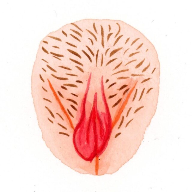
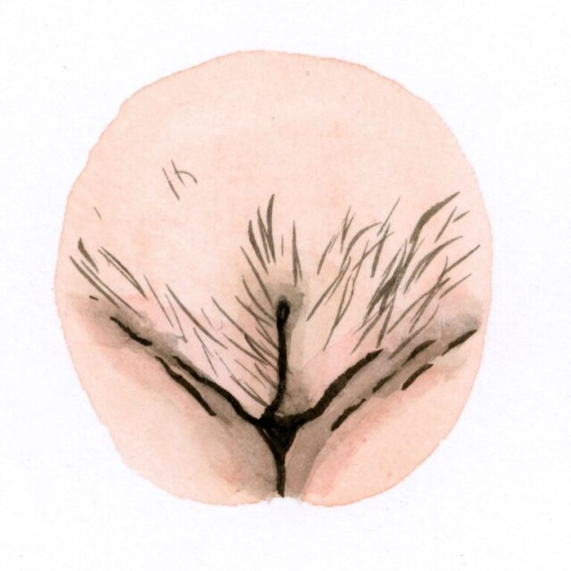
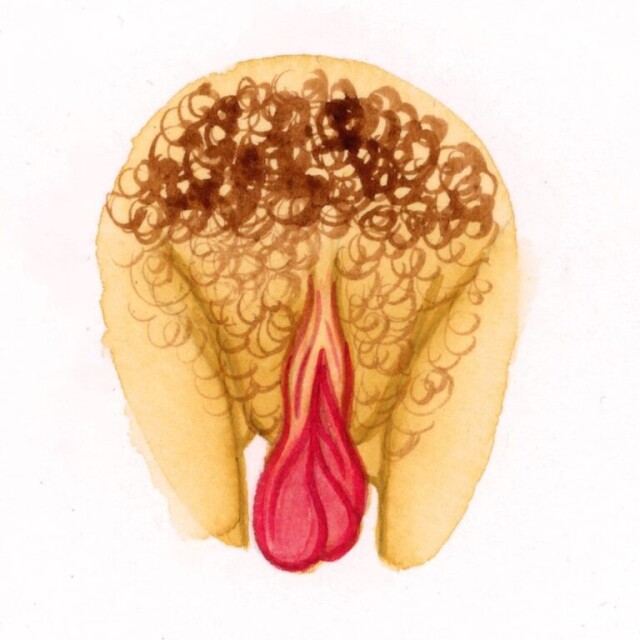
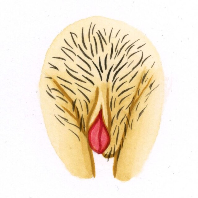
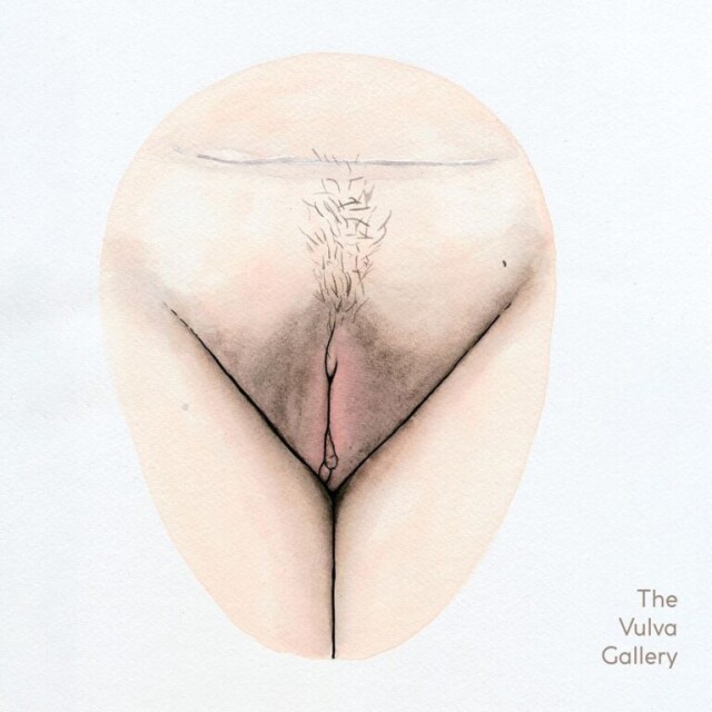
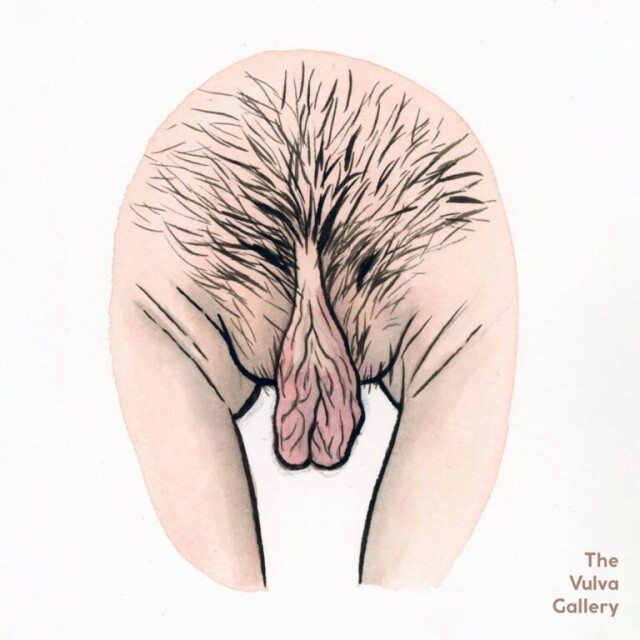
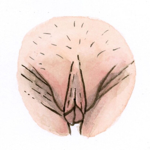
Анатомия клитора
Клитор расположен ниже лобка. Он содержит две эректильные сосудистые ткани, называемые кавернозными телами. Эти ткани располагаются с обеих сторон и окружены оболочкой. Тонкий эпидермис покрывает клитор. Он покрыт многочисленными чувствительными нервами и рецепторами.
Размер клитора остается маленьким до полового созревания и резко увеличивается после. Он состоит из трех важных компонентов, а именно: головки клитора, двух ножек и двух луковиц преддверия. Головка клитора – единственная часть, видимая невооруженным глазом.
Две ножки отходят от головки клитора в виде скобок и углубляются в ткань вульвы. Наконец, две луковицы преддверия простираются по обе стороны от входа во влагалище.
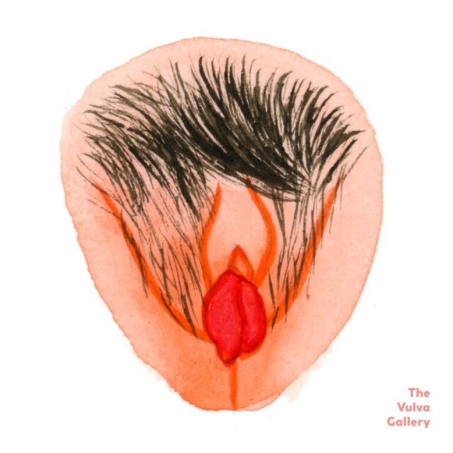
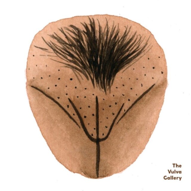
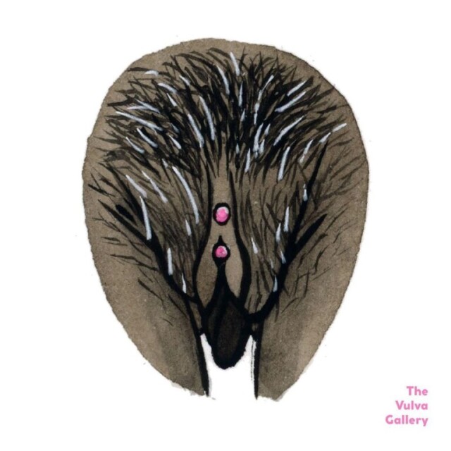
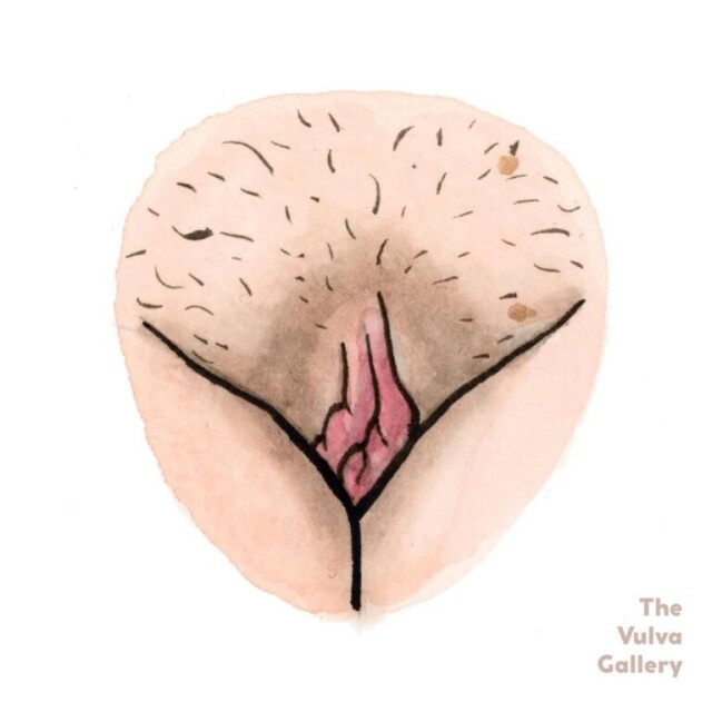
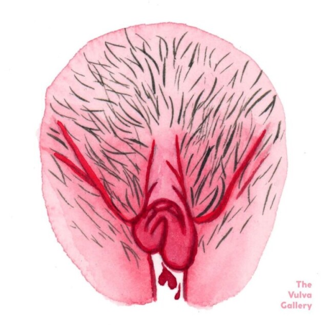
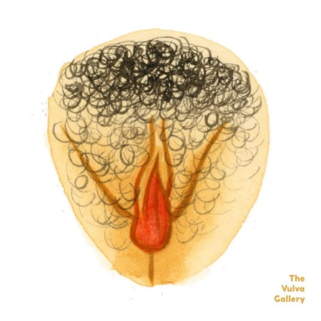
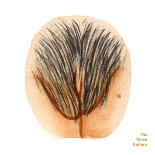
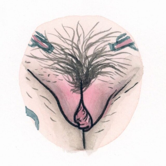
Какого размера средний клитор
Клитор меняется в размерах по мере взросления и изменений в организме. Он увеличивается под действием эстрогенов в репродуктивный период и регрессирует после менопаузы, когда уровень эстрогенов ниже.
Использование гормональной контрацепции, беременность и роды могут привести к некоторому увеличению клитора. Средний размер головки (кончик клитора) составляет от 3 до 10 мм.
У мужчин половой член содержит губчатую ткань, которая набухает, когда наполняется кровью, создавая эрекцию. В клиторе есть похожая губчатая ткань. Когда женщина испытывает сексуальное возбуждение, эта ткань наполняется кровью, и клитор увеличивается.
Клитор снабжён защитной оберткой, называемой капюшоном клитора. Это складка кожи, которая в повседневной жизни накинута на него. Кончик клитора, называемый головкой, густо усеян нервными окончаниями. Большинство медицинских специалистов считают, что оргазм возможен только при стимуляции головки.
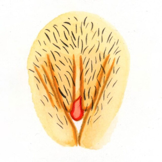
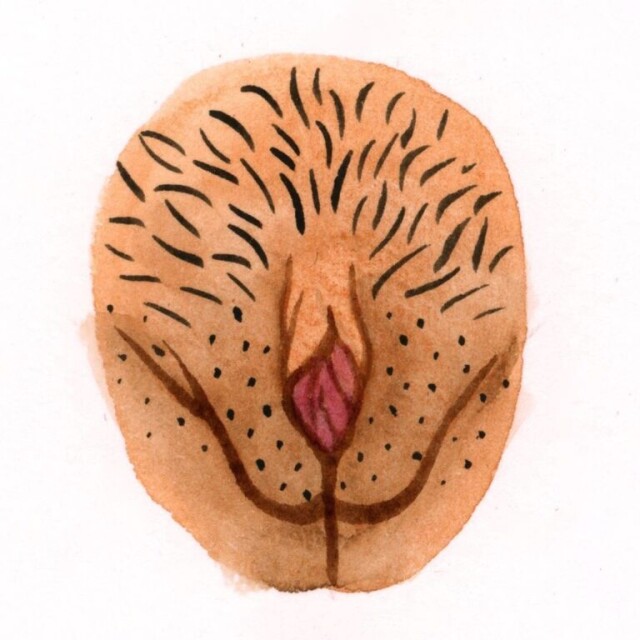
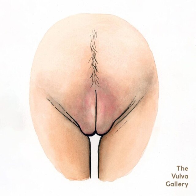
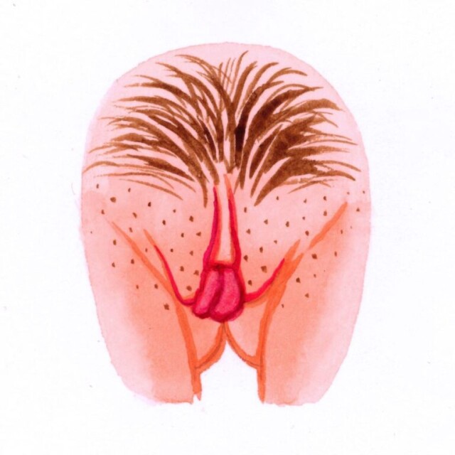
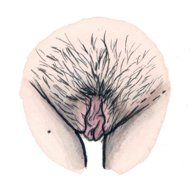
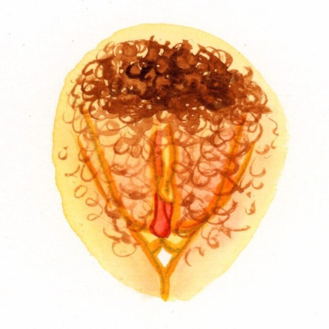
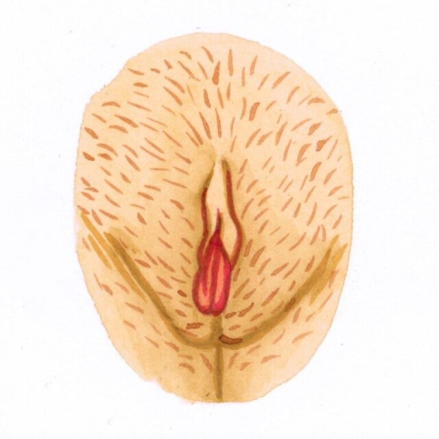
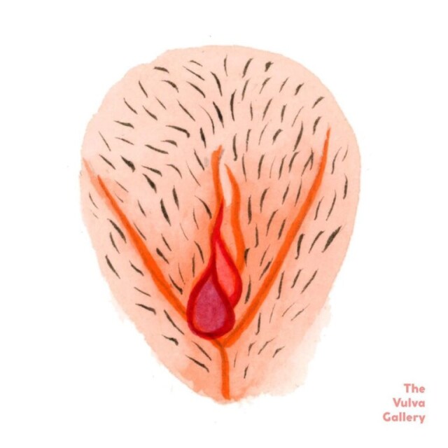
7000
Как работает клитор
Клитор, по-видимому, выполняет сексуальную функцию только у людей, и обычно считается, что он является важным компонентом женского оргазма.
Историки предполагают, что когда-то стимуляция клитора могла быть необходима, чтобы вызвать овуляцию, но в настоящем и недавнем прошлом это определённо не так.
Удивительно, но в клиторе более 8000 нервных окончаний – вдвое больше, чем во всём половом члене, – что объясняет, почему клитор такой чувствительный.
Во время сексуальной активности стимуляция этой области вызывает усиление удовольствия. По мере нарастания возбуждения клитор наполняется кровью, удваивается в размерах и становится эрегированным. Мышечные сокращения происходят в более глубоких частях клитора.

При оргазме кровь выбрасывается из капюшона клитора и заполняет область вокруг. Женский мозг также переполняется гормонами дофамином и окситоцином, что приводит к тому, что женщина одновременно чувствует приподнятое настроение, расслабление и кайф.
Как найти клитор
Если ты – девушка, и не знаешь, где найти свой клитор, не волнуйся. Просто следуй нашему удобному руководству:
- Почему бы не взять зеркало и не посмотреть между ног? Это твоё тело и всё, что есть на нём – абсолютно нормально. Так что не паникуй по поводу того, что ты увидишь;
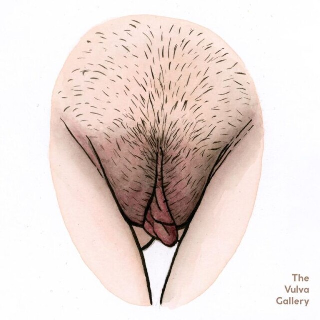
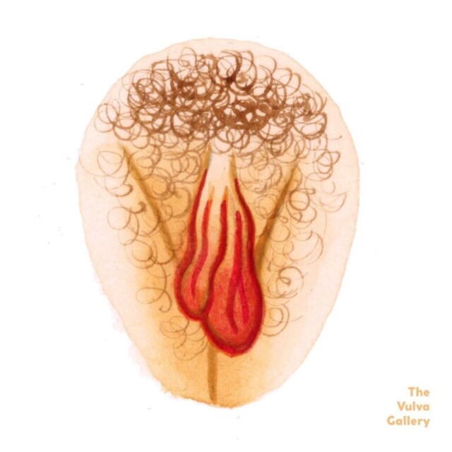
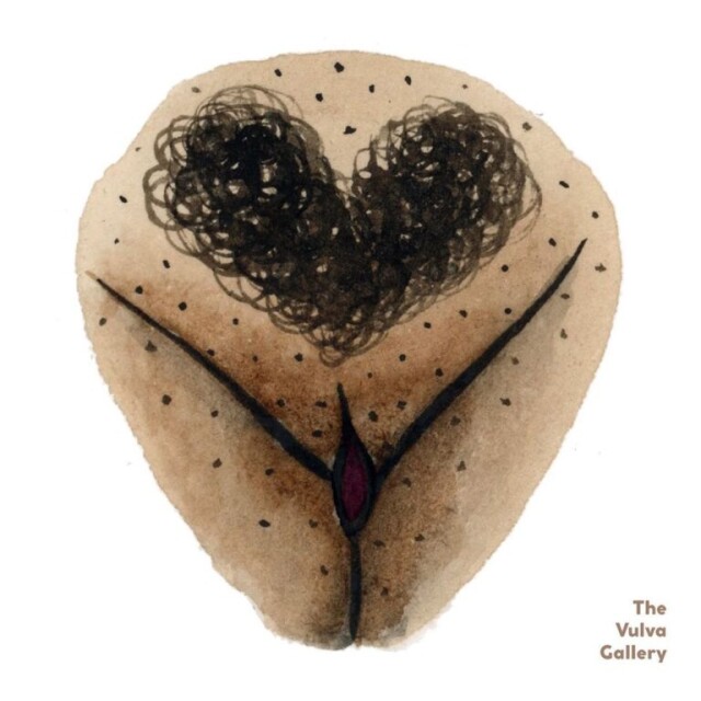
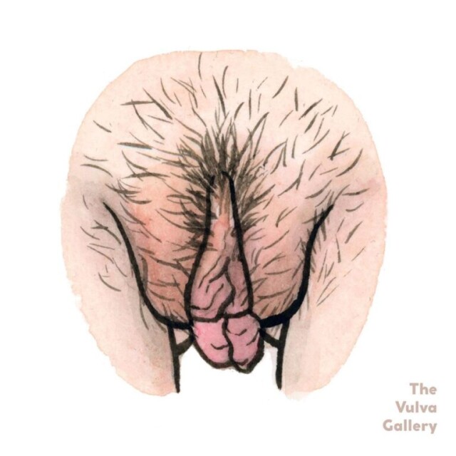
- В центре ты увидишь небольшой лоскут розовой кожи, немного треугольной формы. Это капюшон клитора;
- Одной рукой помести указательный и средний пальцы по обе стороны от этого кожного лоскута. Затем широко раздвинь их, и твой клитор появится перед тобой. Да, это он! Этакая розоватая капля, которая гордо выделяется на фоне всего остального.
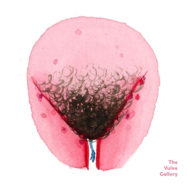
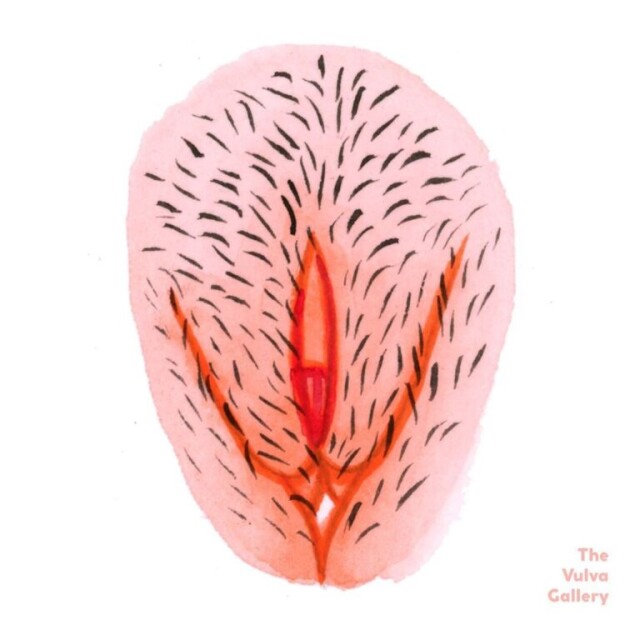
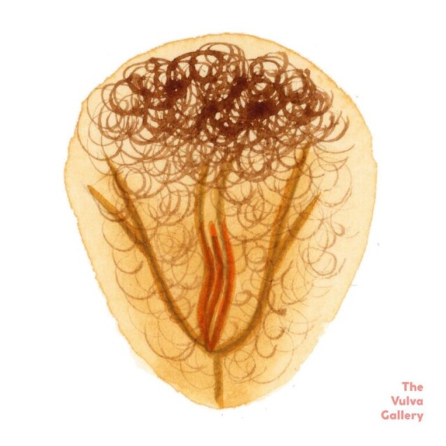
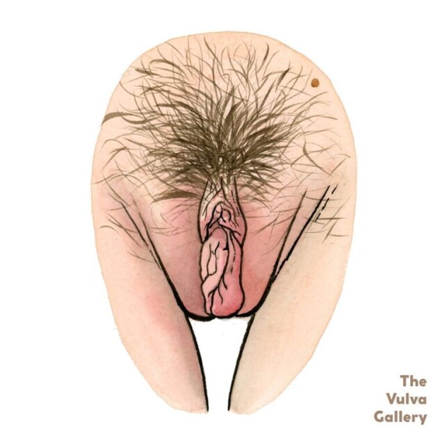
9000
Войны оргазмов
Уже давно ведутся споры о том, что доставляет наибольшее удовольствие: вагинальный оргазм, или оргазм точки G, или клиторальный оргазм. На самом деле был сделан вывод, что только 30% женщин когда-либо испытывают вагинальный оргазм, тогда как клиторальный оргазм может быть достигнут всеми женщинами.
Исследования утверждают, что женский оргазм для большинства с помощью одного лишь проникновения – недостаточен и обязательно нужна стимуляция клитора.
Ещё ряд исследований показывает, что никаких разных видов оргазма не существует. Весь спектр ощущений сосредоточен на нервных окончаниях клитора: неважно, в каком месте при этом происходит стимуляция.
Как достичь оргазма
Известно, что оргазм более вероятен у зрелых и образованных женщин. Вероятно, это потому, что они лучше знают свое тело, а также более уверенно говорят своим сексуальным партнёрам, чего они хотят.
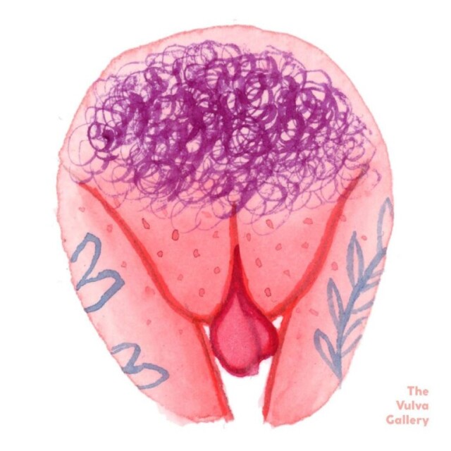
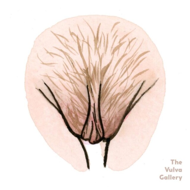
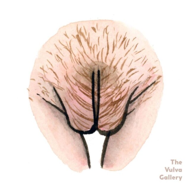
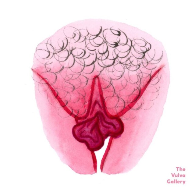
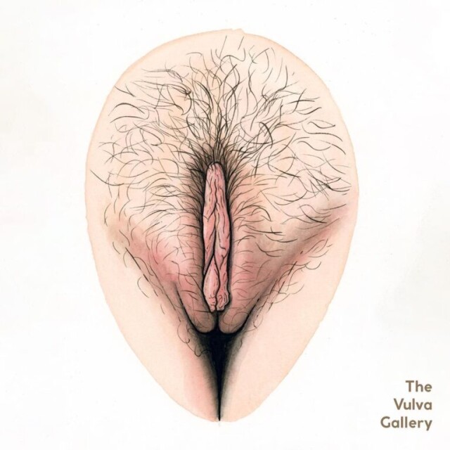
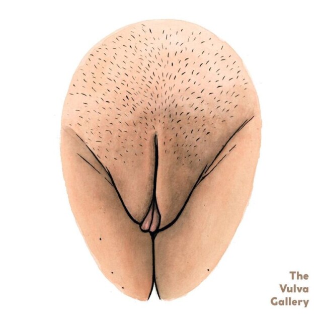
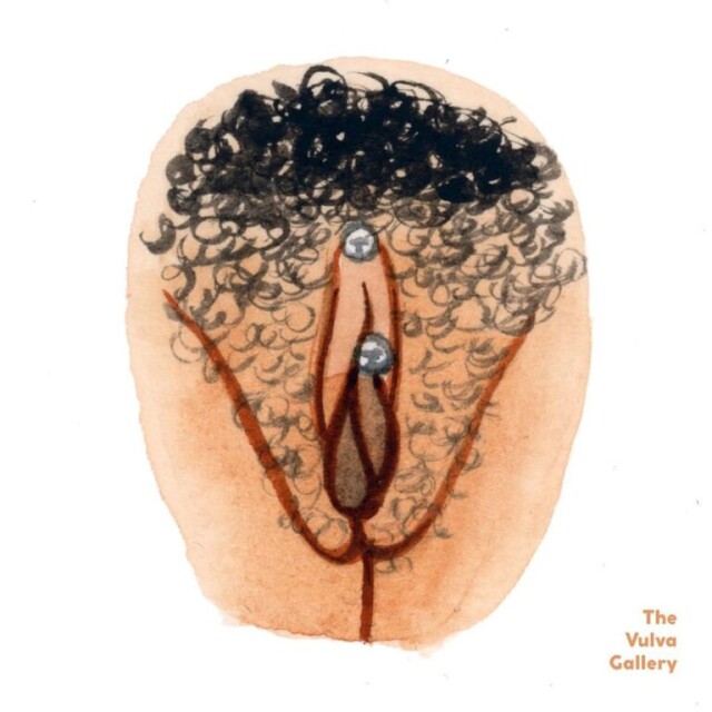
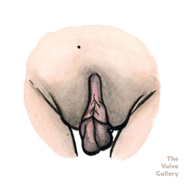
Вот, что важно знать и делать, чтобы испытать оргазм:
- Исследуй своё тело. Убедись, что ты знаешь, где находятся твои эрогенные зоны и как тебе нравится, когда к ним прикасаются. Да – мастурбировать;
- Затем убедись, что ты общаешься с партнёром по поводу секса. Это нормально планировать, обсуждать, договариваться о том, что вам нравится и не нравится. Некоторые пары находят разговоры о сексе эротичными сами по себе. Также важно чувствовать себя расслабленно;
- Если ты не кончаешь во время секса, постарайся не расстраиваться и не унывать. Как и в большинстве вещей, практика ведёт к совершенству.
Понятно, что негативный прошлый опыт, например, такой как сексуальное насилие, является исключением. Если это случилось с тобой, тебе может понадобиться дополнительная психологическая поддержка и помощь.
Это нужно чтобы достичь того уровня, когда ты чувствуешь себя достаточно комфортно, чтобы наслаждаться сексом с понимающим партнёром.
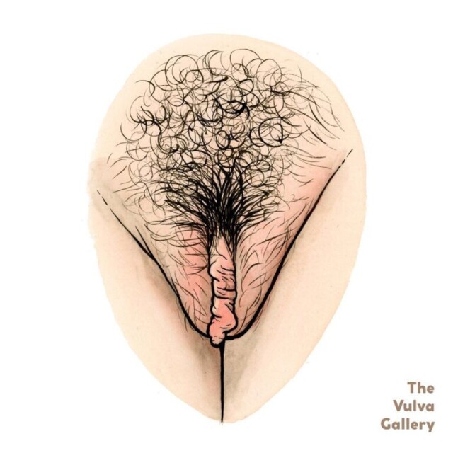
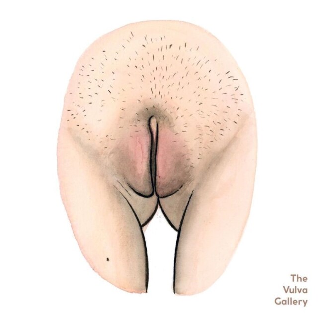
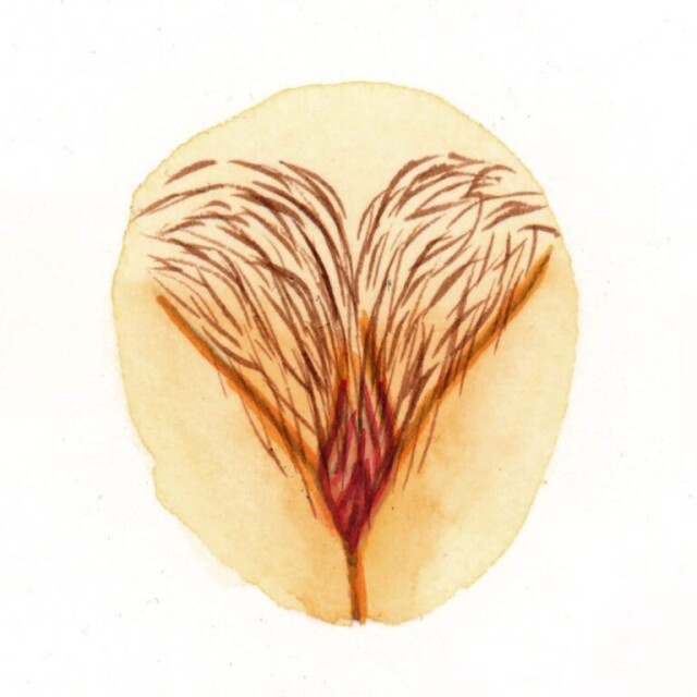
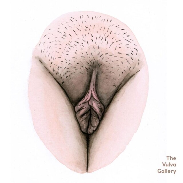
10 интересных фактов о клиторе
О клиторе было сказано уже достаточно, но есть некоторые вещи, о которых вы, возможно, не задумывались.
И прежде чем мы перейдём к фактам, вы должны иметь в виду, что крошечная пуговица в верхней части входа во влагалище, которую ошибочно называют клитором, на самом деле является головкой клитора.
Прочтите это ещё раз – головкой.
Это просто видимая часть клиторальной сети.
Большая часть клитора находится полностью внутри. То, что вы видите, – это лишь «верхушка айсберга».
С учётом сказанного, перейдём к самим фактам.
- Верхняя губа рта женщины и клитор – связаны.
Верхняя губа женщины является одной из эрогенных зон на её теле. Это происходит из-за особого сенсорного нерва под верхней губой, который соединяет её с клитором. Мягкие и нежные поцелуи и посасывания верхней губы женщины безумно возбуждают её.
- Существует также прямая связь между грудью и клитором.
Вы этого, скорее всего, тоже не знали. У вашей груди (или груди вашей женщины) есть прямая горячая линия к клитору. Известно, что женская грудь содержит те же нервные окончания, что и клитор.
Вот почему женщинам приятно, когда их грудь и особенно соски ласкают и сосут.
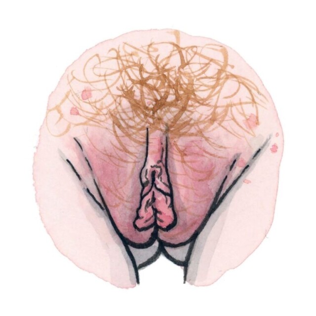
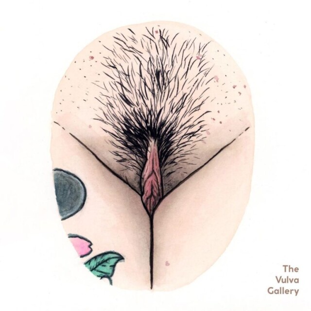
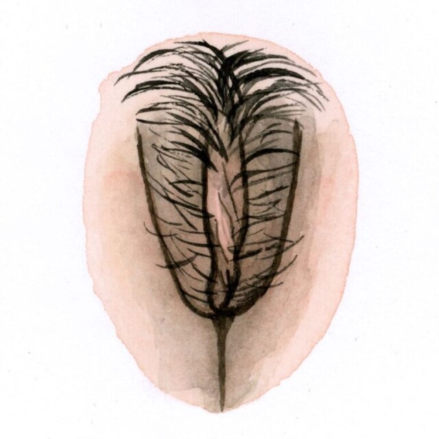
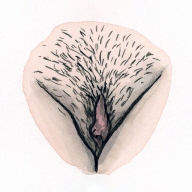
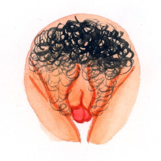
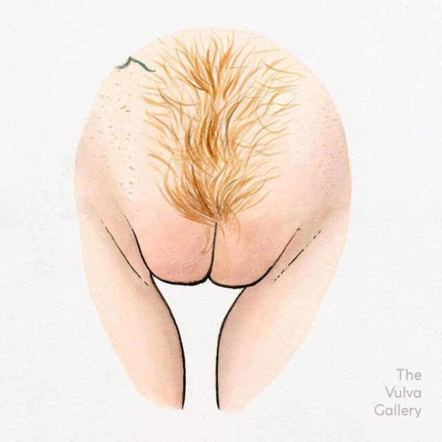
Знаете ли вы, что женщина может испытывать «грудной» оргазм, если её грудь правильно стимулируется?
Да!
Таким образом вместо того, чтобы пренебрегать этими великолепными «двойняшками» и прокладывать себе путь к оргазму, будут оценены небольшие ласки и сосание груди до и во время секса.
- Клитор и анус связаны.
Да, снова связь. Клитор имеет слои мышц, которые связаны с мышцами, окружающими анус. И это одна из причин, почему анальная стимуляция доставляет женщинам удовольствие.
- Корни клитора – точка G.
Есть ли у женщин точка G? Да… Нет… Неважно.
Важно только то, что у них есть клитор. И не только видимый снаружи, но и тот, что находится внутри области таза. Клитор состоит из трёх частей – головки, стержня и ножек (или корней). А ножки перекрещивают губку уретры, где находится «точка G».
Другими словами, передняя стенка влагалища связана с внутренними частями клитора. И стимуляция этой области вызывает более сильный оргазм, отличный от клиторального оргазма.
Исследования показывают, что точка G является частью клиторальной сети. И оргазм точки G, как и все женские оргазмы, является клиторальным оргазмом.
- Клитор вызывает другие оргазмы.
Итак, сколько видов оргазмов может испытать женщина? По подсчётам – около 7.
Эксперты обнаружили, что все оргазмы являются клиторальными, однако все они ощущаются по-разному. Клитор состоит из нескольких частей (как видимых, так и невидимых внутри области влагалища), которые простираются по всей области таза.
Около 8000 клиторальных нервных окончаний взаимодействуют примерно с 15000 других нервных окончаний, «захвативших» всю область таза. А удовольствие и оргазмы, которые женщина испытывает, вызываются 8000 клиторальными нервными окончаниями.
- Клитор окружает вход во влагалище.
Видимая часть клитора – это то, что вы видите над входом во влагалище. Но остальная часть клитора не только уходит глубоко в гениталии, но и окружает вагинальное отверстие.
Когда женщина возбуждается, эректильная ткань клитора набухает. Это ещё одна причина, по которой ей приятно, когда что-то скользит туда-сюда внутри её влагалища.
- Части клитора также лежат по обе стороны от вагинального канала.
По этой причине стимуляция стенок вагинального канала пальцами доставляет женщине удовольствие.
- Корни клитора проходят параллельно большим половым губам (они же внешние вагинальные губы).
И это ещё одна причина, по которой вам следует стимулировать большие половые губы (две внешние складки вульвы) во время прелюдии и орального секса.
- Малые половые губы имеют внешнее соединение с головкой клитора.
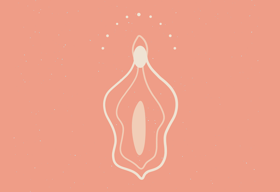
Малые половые губы – это губы, которые прячутся за большими половыми губами (внешними). Хотя иногда они выступают за внешние губы у некоторых женщин.
Благодаря соединению с головкой и капюшоном клитора, облизывание складок внутри, снаружи и вокруг складок доставляет огромное удовольствие.
- Он встаёт так же, как пенис.
Если вы ещё не знали, клитор (в частности, стержень) выпрямляется так же, как пенис. Каким, казалось бы, маленьким ни казался клитор, он содержит эректильную ткань (точно так же, как пенис), которая становится эрегированной и напрягается, когда женщина возбуждается.
Клитор наполняется кровью и становится толще из-за возбуждения. И стимуляция на этом этапе подготовит женщину к интенсивному оргазму в кратчайшие сроки.
Как правильно ласкать себя
Мастурбация сильно недооценивается. Это самый лёгкий способ изучить своё тело, получить дозу гормонов счастья и быть более раскрепощённой со своим партнёром. И вот, какими способами можно получить клиторальный оргазм.
- Попробуй чувственную стимуляцию.
Чувственная стимуляция – это способ дразнить и разбудить тело, так что возбуждение нарастает и нарастает, пока оно не будет готово перелиться в оргазм или в желание проникновения. Она предлагает использовать медленные и чувственные прикосновения ко всему телу (а не только к клитору) и начинать снаружи внутрь.
Клитор и влагалище должны быть последними в порядке стимуляции. Она предлагает двигаться от подмышек к груди (не касаясь сосков), к внутренней поверхности бедер.
Как только ты дойдёшь до гениталий, начни с внешних половых губ и двигайся внутрь.
- Попробуй метод «знак мира».
Если у тебя очень чувствительный клитор, непосредственное прикосновение к нему может показаться чрезмерной стимуляцией.
Для женщин с ультрачувствительными клиторами подними два пальца вверх, как будто вы делаете знак мира (указательный и средний палец немного разведи в стороны), и нажмите за пределами клитора, на внешних половых губах.
Оттуда ты можешь либо двигать пальцами вверх и вниз (представь, что ты играешь на двух клавишах на пианино), либо мягко массировать пальцами вместе, а затем врозь. Оба эти движения могут стимулировать твой клитор без прямого контакта.
- Постарайся стимулировать весь клитор, а не только его видимую внешнюю часть.
Твой клитор подобен айсбергу – под поверхностью гораздо больше нервов, и та часть, которую ты можешь увидеть и потрогать, на самом деле является лишь небольшой его частью. Твой клитор на простирается вокруг входа во влагалище и может быть сравним с рычагом.
Помассируй половые губы и область прямо над поперечными ножками клитора для ещё большей стимуляции. Ты также можешь расширить свои прикосновения ещё дальше и массировать вход во влагалище, промежность (между влагалищем и анусом), внутреннюю поверхность бёдер и анус.
- Используй лёгкое давление.
Кожа, покрывающая твой клитор, – это твой клиторальный капюшон, и, по сути, он похож на крайнюю плоть. Ты также можешь использовать капюшон клитора во время сеансов мастурбации.
Осторожно зажми кожу между большим и указательным пальцами, и поэкспериментируй с давлением, пока не найдёшь то, что доставляет тебе удовольствие. Оттуда ты можешь попробовать разные движения, например, двигать рукой или пальцами вверх и вниз.
- Попробуй диагональные движения.
«Потирать» клитор – это довольно расплывчатое указание, поэтому иногда может быть полезно точно знать, что делать и как двигать пальцами или руками.
С помощью двух-трёх пальцев нежно потри клитор вперед и назад диагональными движениями, увеличивая скорость и интенсивность по своему желанию. Это может быть здорово, если у тебя чувствительный клитор или ты просто хочешь что-то изменить.
- Используй смазку.
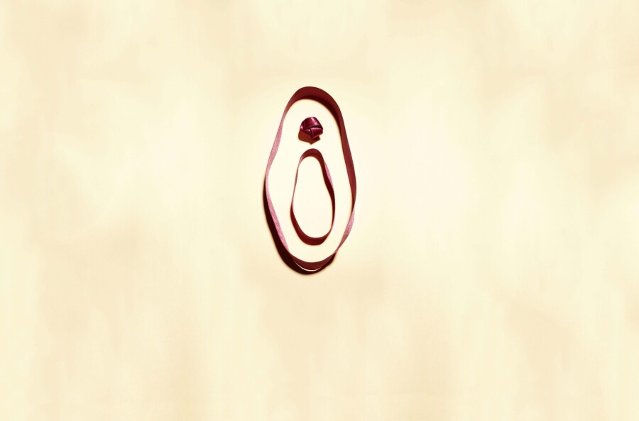
Нанеси на пальцы каплю смазки и нежно помассируй ею клитор. Ты также можешь положить пальцы по обе стороны от клитора и медленно двигаться вверх и вниз по внутренним половым губам, то есть по складкам кожи, непосредственно окружающим клитор.
Смазка позволяет тебе замедлиться и по-настоящему сосредоточиться на плавных, непрерывных движениях, чтобы ты могла понять, что тебе нравится. Не все любят трение.
- Попробуй круговые движения.
Некоторые женщины считают, что постоянство, а не увеличение темпа, – это то, что их раздражает. Чтобы проверить эту теорию, используй один или два пальца, чтобы сделать круги вокруг или на верхушке твоего клитора.
Ты можешь увеличивать скорость, но как только ты найдёшь то, что тебе нравится, попробуй поработать несколько минут и посмотреть, что произойдёт.
- Попробуй лёгкие постукивания.
Вишенка на торте: по-прежнему спокойно, но сейчас сделай это постукивающим движением. Это может утомить твою руку через некоторое время, но позволяет тебе играть с давлением.
И определи для себя: тебе нравятся постукивания, которые кажутся немного более тяжелыми, как растирание, или более лёгкими, как прохладный океанский бриз, который доставляет тебе бомбические оргазмы.
- Попробуй удвоить время.
Попробуй использовать обе руки. Одна рука медленно обводит вход во влагалище, не проникая внутрь, в то время как другая быстрее рисует круги на клиторе. Увеличение разнообразия ощущений может быть именно тем, что тебе нужно.
- Попробуй на животе.
Положите подушку под живот и устройся поудобнее. Если ты из тех, кто любит тереть клитор о поверхность, то такой вариант отлично подойдёт. Ты можешь посмотреть, что происходит, когда мастурбируешь лицом вниз на своей кровати.
Таким образом, ты по-прежнему можешь использовать свои руки, но также можешь свободно тереться о матрас или подушку, чтобы получить больше удовольствия.
- Попробуй вибратор.
В конце концов, вибраторы – отличный вариант для женщин, у которых устают руки или, будем честны, если у тебя длинные ногти. В основном, вибраторы для всех. Они ТОП для экспериментов, потому что большинство игрушек в наши дни различаются как по скорости, так и по характеру вибрации.
Вы можешь найти звёздный список вибраторов, ориентированных на клитор в любом секс-шопе. Просто убедись, что его аккумулятор полностью заряжен, прежде чем начать его использовать. Тебе ведь не захочется прерывать удовольствие.
- Используй партнёра.
Да, этот вариант тоже достоин быть здесь. Совместно с партнёром вы получаете ещё больше возможностей стимулировать твой клитор.
Не входя в тебя, партнёр может ублажать тебя орально, используя разные движения языка или подключая пальцы. Он также может гладить и массировать твои половые губы, пока ты сосредоточена на клиторе.
Партнёр может ласкать тебя головкой члена без непосредственного проникновения, что будет усиливать возбуждение и желание. А такие действия быстро приведут к оргазму. Ты также можешь сесть на него сверху и тереться, как о любой другой предмет. Или сесть на лицо.
Теперь вы знаете немного больше о клиторе, его строении и способах достижения оргазма. Девушки, не стесняйтесь своего тела и позвольте себе получать удовольствие. Мужчины, будьте внимательны к своим партнёршам и не «теряйте» клитор.
И это все о нем
«Камасутра» сравнивает женские половые органы с таинственной долиной, о которой мечтает любящий мужчина… Она дивно благоухает, скрывая в своих недрах тайное сокровище – это клитор. У него единственная забота — обеспечивать сексуальную чувствительность. Такова уж особенность этого органа наслаждения.
Располагается клитор под верхним сращением малых половых губ и скрыт крайней плотью. Возбуждаясь, наполняется кровью и становится самой чувствительной точкой женского тела. Выступающая из-под крайней плоти часть клитора — лишь его головка, видимая часть, которая составляет всего 4 процента эректильной системы клитора.
Головка – венец скрытого внутри сокровища, которое стоит на двух ножках. Весь клитор – губчатая ткань, которая как и пенис у мужчин твердеет при возбуждении, наполняясь кровью. Причем приток крови к половым органам женщины существенно больше, чем к пенису у мужчины, а заметна женская эрекция гораздо слабее.
Размер клитора неважен. Значительная часть женщин вообще не обнаруживает его у себя. Тому есть несколько причин. Одна из них решается только хирургически, надрезом освобождая клитор из плена, другая причина — минимальный размер образования. Повода для расстройства нет, ведь все вокруг клитора пронизано нервными окончаниями, вызывающими оргазм.
Найти клитор просто: разведи бедра, осторожно раздвинь большие и малые половые губы, найди в месте их сращивания складку. Это крайняя плоть клитора, которую часто называют капюшоном. Приподними его кверху, и увидишь маленькую розовую выпуклость-пуговку. Это и есть клитор. В момент прикосновения к ней ты испытаешь сладострастное ощущение.
Лучше всего в момент интима доверить клитор рту. Язык и губы замечательно приспособлены для ласк клитора: они мягки и влажны, а их движения ловки и точны. У некоторых женщин клитор плохо раздражается при совокуплении. Вот почему, вводя половой член во влагалище женщины, мужчина ни в коем случае не должен прекращать ласки на клиторе.
Клитор не всегда возбуждается сразу. И это заметно по отсутствию секреторной жидкости, выделяющейся из половых органов женщины. Даже если не было уделено достаточно внимания любовной предварительной игре, а женщина находится в настроении, благоприятствующему половому акту, то всегда выделяется достаточное количество жидкости и концентрируется в области клитора.
Всем известно, что клитор является женским центром получения сексуального удовольствия. Это своеобразный аналог мужского члена, только на много меньше и на много чувствительней, поскольку имеет в 2 раза больше нервных окончаний. Очень часто девушки остаются неудовлетворенны по элементарной причине мало того, что мужчина не знает как обращаться с этой жемчужиной женского удовольствия, он еще толком и не понимает где расположен клитор. Именно поэтому если вы хотите гарантировано довести девушку до оргазма, вы обязаны понимать где находится клитор у девушки, знать как его находить и как с ним грамотно обращаться.
Расположение клитора у девушек
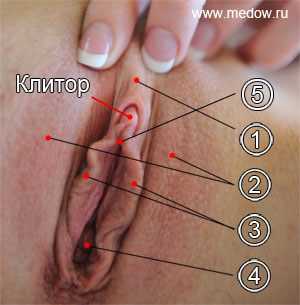 Чтоб выяснить его расположение давайте посмотрим на фото слева и разберемся, что там изображено.
1) Капюшон (крайняя плоть),
2) Большие половые губы,
3) Малые половые губы,
4) Вход во влагалище,
5) Уздечка.
Все что вы видите на этой картинке называется женские гениталии. А клитор, а точней его головка – это самая чувствительная их часть. Головка клитора находится сразу над верхней точкой скрещения малых половых губ и представляет собой маленькую горошинку. На фото, как вы видите, она подписана.
Следует также понимать, что сам по себе клитор на много больше. Визуально видна всего лишь его головка, а основное тело его находится под кожей, но об этом ниже.
Видимая часть состоит из головки, капюшона (1) и уздечки (5). Самой чувствительной является головка и чем она меньше тем более чувствительна. Так, чуть выше малых половых губ есть небольшая складка кожи, которая называется капюшоном (1) или крайней плотью. Капюшон выполняет защитную функцию, оборачивая и защищая от лишнего трения сверхчувствительную головку. Капюшон является аналогом крайней плоти у мужчин. Сама головка выглядит как маленькая розовая выпуклость размером с горошину. Она не всегда видна, поэтому для того, чтобы ее увидеть, нужно просто аккуратно положить палец на капюшон и сделать движение пальцем по направлению к лобку. Стянув таким образом капюшон, вы увидите головку. Также стоит сказать о уздечке (5) потому как она также очень чувствительна и ее стимуляции также очень эффективна в процессе возбуждения девушки. Уздечка это небольшой центральный участок перехода малых губ в головку клитора.
Строение клитора и его эрогенные зоны
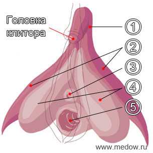 Не смотря на то, что внешне клитор кажется достаточно простым и маленьким органом, на самом деле он на много больше и имеет куда более сложное строение. Все дело все в том, что большая его часть уходит под кожу. Давайте опять посмотрим на картинку.
1) Тело (может достигать 10 см в длину),
2) Ножки,
3) Уретра или мочеиспускательный канал,
4) Луковица преддверия влагалища,
5) Вход во влагалище.
В целом клитор имеет форму перевернутой буквы V. Его ножки (2) охватывают вход во влагалище, так что во время секса стимулируется не только само влагалище, но и клитор тоже, по крайней мере, невидимая его часть. Благодаря этому получаемое удовольствие в немалой степени усиливается. Хотя стоит признать, что головка все же является гораздо более чувствительной.
Что касается наиболее чувствительных зон после головки клитора, то здесь все очень неоднозначно. Дело в том, что у каждой девушки различные его части могут быть в разной степени чувствительными к стимуляции. Тем не менее, можно выделить 2 основных эпицентра наслаждения:
- Сама головка,
- Уздечка,
Как найти клитор?
Существует 2 основных способа как можно найти клитор у девушки: визуальный и тактильный. Они оба хороши, хотя в идеале лучше использовать их одновременно. Тем не менее, в определенных условиях это невозможно, так что будет не лишним знать в подробностях о каждом из них.
- Визуальный способ для начала девушка должна лечь на спину и немного раздвинуть ноги, между которыми вы должны разместиться. Под лобком вы увидите вагину (см первую картинку), наружная часть которой состоит из больших(2) и малых(3) половых губ, входа во влагалище(4) и капюшона (1), который находится в самой верхней части. Прежде всего, при помощи пальцев рук следует сначала немного раздвинуть большие половые губы чтоб вы четко видели ее малые губы. Так вот та точка в которой сходятся 2 малые губы и является клитором (его головкой). Иногда в силу того, что форма половых органов у каждой девушки неповторима, внешнее расположение клитора и его размеры также могут отличатся. Так например у одной девушки он может быть более выразителен за счет размеров и не слишком скрыт под капюшоном, а у другой наоборот для того чтоб его обнаружить нужно пальцем отодвигать крайнюю плоть. В общем в каждом конкретном случае он может выглядеть по разному.
- Тактильный способ в том случае, если близость происходит в условиях полумрака или полной темноты, либо когда выбранная позиция ограничивает осмотр, визуальный метод поиска становится неэффективным. И в такой ситуации будет гораздо легче нащупать клитор. Для этого обязательно смочите палец слюной и положите его на лобок. А именно в точку, где сходятся большие половые губы. Затем едва надавив, волнообразными движениями из стороны в сторону спускайтесь к входу во влагалище, а точней к точке, где сходятся малые половые губы . Вы почувствуете как под пальцем у вас проскакивает словно тоненькая веревочка. Это и есть тело клитора, которое уходит под кожу и чем сильней возбуждена девушка тем выразительней становится она (эта жилка под пальцем). Спускаясь по этой веревочке вниз в сторону входа во влагалище вы почувствуете утолщение в виде складки кожи вот это есть капюшон под которым прячется самая чувствительная часть клитора его головка. Именно это место самое важное для стимуляции и именно то место которое так старательно пытаются найти парни у девушки. Очень важный момент: у всех девушек клитор разный по чувствительности, поэтому первые прикосновения к нему должны быть очень и очень нежными на случай того что у девушки он может быть как раз очень чувствительным. Более подробно о типах клитора читайте в статье клитор. Чтоб сразу исключить нелепые ситуации, советую сразу девушку попросить дать вам знать если ей вдруг станет неприятно. А то знаете как часто бывает:
Ласкаешь клитор пальцем, а девушка стонет и вся извивается. Вы думаете что она балдеет от ваших ласк, а она извивается и стонет от боли и не может дождаться того момента когда вы прекратите.
Надеюсь, что после прочтения этой статьи ни у кого из парней не останется вопросов по поводу того, где находится клитор и как его найти. Твердо зная это вы сможете на много лучше делать кунилингус или просто эротический массаж клитора.
Тест: Не изменяет ли ваша вторая половина?
Вы подозреваете, что ваш партнёр вас обманывает? Пройдите тест и узнайте, может ли вас водить за нос ваша вторая половина.

( 7 оценок, среднее 3.29 из 5 )
| Clitoris | |
|---|---|

The internal anatomy of the human vulva, with the clitoral hood and labia minora indicated as lines. The clitoris extends from the visible portion to a point below the pubic bone. |
|
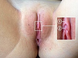
Location of (1) clitoral hood and (2) clitoral glans |
|
| Details | |
| Precursor | Genital tubercle |
| Artery | Dorsal artery of clitoris, deep artery of clitoris |
| Vein | Superficial dorsal veins of clitoris, deep dorsal vein of clitoris |
| Nerve | Dorsal nerve of clitoris |
| Identifiers | |
| Latin | Clitoris |
| MeSH | D002987 |
| TA98 | A09.2.02.001 |
| TA2 | 3565 |
| FMA | 9909 |
| Anatomical terminology
[edit on Wikidata] |
The clitoris ( or ) is a female sex organ present in mammals, ostriches and a limited number of other animals. In humans, the visible portion – the glans – is at the front junction of the labia minora (inner lips), above the opening of the urethra. Unlike the penis, the male homologue (equivalent) to the clitoris, it usually does not contain the distal portion (or opening) of the urethra and is therefore not used for urination. In most species, the clitoris lacks any reproductive function. While few animals urinate through the clitoris or use it reproductively, the spotted hyena, which has an especially large clitoris, urinates, mates, and gives birth via the organ. Some other mammals, such as lemurs and spider monkeys, also have a large clitoris.[1]
The clitoris is the human female’s most sensitive erogenous zone and generally the primary anatomical source of human female sexual pleasure.[2] In humans and other mammals, it develops from an outgrowth in the embryo called the genital tubercle. Initially undifferentiated, the tubercle develops into either a penis or a clitoris during the development of the reproductive system depending on exposure to androgens (which are primarily male hormones). The clitoris is a complex structure, and its size and sensitivity can vary. The glans (head) of the human clitoris is roughly the size and shape of a pea and is estimated to have 8,000, and possibly more than 10,000, sensory nerve endings.[3][4]
Sexological, medical, and psychological debate has focused on the clitoris,[5] and it has been subject to social constructionist analyses and studies.[6] Such discussions range from anatomical accuracy, gender inequality, female genital mutilation, and orgasmic factors and their physiological explanation for the G-spot.[7] Although, in humans, the only known purpose of the clitoris is to provide sexual pleasure, whether the clitoris is vestigial, an adaptation, or serves a reproductive function has been debated.[8] Social perceptions of the clitoris include the significance of its role in female sexual pleasure, assumptions about its true size and depth, and varying beliefs regarding genital modification such as clitoris enlargement, clitoris piercing and clitoridectomy.[9] Genital modification may be for aesthetic, medical or cultural reasons.[9]
Knowledge of the clitoris is significantly impacted by cultural perceptions of the organ. Studies suggest that knowledge of its existence and anatomy is scant in comparison with that of other sexual organs and that more education about it could help alleviate social stigmas associated with the female body and female sexual pleasure, for example, that the clitoris and vulva in general are visually unappealing, that female masturbation is taboo, or that men should be expected to master and control women’s orgasms.[10]
Etymology
The Oxford English Dictionary states that the word clitoris likely has its origin in the Ancient Greek κλειτορίς, kleitoris, perhaps derived from the verb κλείειν, kleiein, “to shut”.[11] Clitoris is also Greek for the word key, “indicating that the ancient anatomists considered it the key” to female sexuality.[12][13] In addition to key, the Online Etymology Dictionary suggests other Greek candidates for the word’s etymology include a noun meaning “latch” or “hook”; a verb meaning “to touch or titillate lasciviously”, “to tickle” (one German synonym for the clitoris is der Kitzler, “the tickler”), although this verb is more likely derived from “clitoris”; and a word meaning “side of a hill”, from the same root as “climax”.[14] The Oxford English Dictionary also states that the shortened form “clit”, the first occurrence of which was noted in the United States, has been used in print since 1958: until then, the common abbreviation was “clitty”.[11]
The plural forms are clitorises in English and clitorides in Latin. The Latin genitive is clitoridis, as in “glans clitoridis”. In medical and sexological literature, the clitoris is sometimes referred to as “the female penis” or pseudo-penis,[15] and the term clitoris is commonly used to refer to the glans alone;[16] partially because of this, there have been various terms for the organ that have historically confused its anatomy.
Structure
Development
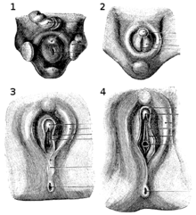
In mammals, sexual differentiation is determined by the sperm that carries either an X or a Y (male) chromosome.[17] The Y chromosome contains a sex-determining gene (SRY) that encodes a transcription factor for the protein TDF (testis determining factor) and triggers the creation of testosterone and anti-Müllerian hormone for the embryo’s development into a male.[18][19] This differentiation begins about eight or nine weeks after conception.[18] Some sources state that it continues until the twelfth week,[20] while others state that it is evident by the thirteenth week and that the sex organs are fully developed by the sixteenth week.[21]
The clitoris develops from a phallic outgrowth in the embryo called the genital tubercle. Initially undifferentiated, the tubercle develops into either a clitoris or penis during the development of the reproductive system depending on exposure to androgens (which are primarily male hormones). The clitoris forms from the same tissues that become the glans and shaft of the penis, and this shared embryonic origin makes these two organs homologous (different versions of the same structure).[22]
If exposed to testosterone, the genital tubercle elongates to form the penis. By fusion of the urogenital folds – elongated spindle-shaped structures that contribute to the formation of the urethral groove on the belly aspect of the genital tubercle – the urogenital sinus closes completely and forms the spongy urethra, and the labioscrotal swellings unite to form the scrotum.[22] In the absence of testosterone, the genital tubercle allows for the formation of the clitoris; the initially rapid growth of the phallus gradually slows and the clitoris is formed. The urogenital sinus persists as the vestibule of the vagina, the two urogenital folds form the labia minora, and the labioscrotal swellings enlarge to form the labia majora, completing the female genitalia.[22] A rare condition that can develop from higher than average androgen exposure is clitoromegaly.[23]
Gross anatomy and histology
General

Created by Helen O’Connell using MRI, the first 3D image of a clitoris in an erect state with the adjacent organs of the uterus and urinary bladder
Clitoris; deep dissection
The clitoris contains external and internal components. It consists of the glans, the body (which is composed of two erectile structures known as the corpora cavernosa), and two crura (“legs”). It has a hood formed by the labia minora (inner lips). It also has vestibular or clitoral bulbs. The frenulum of the clitoris is a frenulum on the undersurface of the glans and is created by the two medial parts of the labia minora.[24] The clitoral body may be referred to as the shaft (or internal shaft), while the length of the clitoris between the glans and the body may also be referred to as the shaft. The shaft supports the glans, and its shape can be seen and felt through the clitoral hood.[25]
Research indicates that clitoral tissue extends into the vagina’s anterior wall.[26] Şenaylı et al. said that the histological evaluation of the clitoris, “especially of the corpora cavernosa, is incomplete because for many years the clitoris was considered a rudimentary and nonfunctional organ.” They added that Baskin and colleagues examined the clitoris’s masculinization after dissection and using imaging software after Masson chrome staining, put the serial dissected specimens together; this revealed that the nerves of the clitoris surround the whole clitoral body (corpus).[27]
The clitoris, vestibular bulbs, labia minora, and urethra involve two histologically distinct types of vascular tissue (tissue related to blood vessels), the first of which is trabeculated, erectile tissue innervated by the cavernous nerves. The trabeculated tissue has a spongy appearance; along with blood, it fills the large, dilated vascular spaces of the clitoris and the bulbs. Beneath the epithelium of the vascular areas is smooth muscle.[28] As indicated by Yang et al.’s research, it may also be that the urethral lumen (the inner open space or cavity of the urethra), which is surrounded by a spongy tissue, has tissue that “is grossly distinct from the vascular tissue of the clitoris and bulbs, and on macroscopic observation, is paler than the dark tissue” of the clitoris and bulbs.[29] The second type of vascular tissue is non-erectile, which may consist of blood vessels that are dispersed within a fibrous matrix and have only a minimal amount of smooth muscle.[28]
Glans and body

A partially exposed clitoral glans, which cannot be fully exposed due to a mild case of adhesions to the clitoral hood
Highly innervated, the glans exists at the tip of the clitoral body as a fibro-vascular cap[28] and is usually the size and shape of a pea, although it is sometimes much larger or smaller. The clitoral glans, or the entire clitoris, is estimated to have 8,000, and possibly 10,000 or more, sensory nerve endings.[3][4] Research conflicts on whether or not the glans is composed of erectile or non-erectile tissue. Although the clitoral body becomes engorged with blood upon sexual arousal, erecting the clitoral glans, some sources describe the clitoral glans and labia minora as composed of non-erectile tissue; this is especially the case for the glans.[16][28] They state that the clitoral glans and labia minora have blood vessels that are dispersed within a fibrous matrix and have only a minimal amount of smooth muscle,[28] or that the clitoral glans is “a midline, densely neural, non-erectile structure”.[16]
Other descriptions of the glans assert that it is composed of erectile tissue and that erectile tissue is present within the labia minora.[30] The glans may be noted as having glandular vascular spaces that are not as prominent as those in the clitoral body, with the spaces being separated more by smooth muscle than in the body and crura.[29] Adipose tissue is absent in the labia minora, but the organ may be described as being made up of dense connective tissue, erectile tissue and elastic fibers.[30]
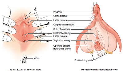
Structures of the vulva, including external and internal parts of the clitoris

The clitoral body forms a wishbone-shaped structure containing the corpora cavernosa – a pair of sponge-like regions of erectile tissue that contain most of the blood in the clitoris during clitoral erection. The two corpora forming the clitoral body are surrounded by thick fibro-elastic tunica albuginea, literally meaning “white covering”, connective tissue. These corpora are separated incompletely from each other in the midline by a fibrous pectiniform septum – a comblike band of connective tissue extending between the corpora cavernosa.[27][28]
The clitoral body extends up to several centimeters before reversing direction and branching, resulting in an inverted “V” shape that extends as a pair of crura (“legs”).[31] The crura are the proximal portions of the arms of the wishbone. Ending at the glans of the clitoris, the tip of the body bends anteriorly away from the pubis.[29] Each crus (singular form of crura) is attached to the corresponding ischial ramus – extensions of the copora beneath the descending pubic rami.[27][28] Concealed behind the labia minora, the crura ends with attachment at or just below the middle of the pubic arch.[N 1][33] Associated are the urethral sponge, perineal sponge, a network of nerves and blood vessels, the suspensory ligament of the clitoris, muscles and the pelvic floor.[28][34]
There is no identified correlation between the size of the clitoral glans or clitoris as a whole, and a woman’s age, height, weight, use of hormonal contraception, or being post-menopausal, although women who have given birth may have significantly larger clitoral measurements.[35] Centimeter (cm) and millimeter (mm) measurements of the clitoris show variations in its size. The clitoral glans have been cited as typically varying from 2 mm to 1 cm and usually being estimated at four to five mm in both the transverse and longitudinal planes.[36]
A 1992 study concluded that the total clitoral length, including glans and body, is 16.0 ± 4.3 mm (0.63 ± 0.17 in), where 16 mm (0.63 in) is the mean and 4.3 mm (0.17 in) is the standard deviation.[37] Concerning other studies, researchers from the Elizabeth Garrett Anderson and Obstetric Hospital in London measured the labia and other genital structures of 50 women from the age of 18 to 50, with a mean age of 35.6., from 2003 to 2004, and the results given for the clitoral glans were 3–10 mm for the range and 5.5 [1.7] mm for the mean.[38] Other research indicates that the clitoral body can measure 5–7 centimetres (2.0–2.8 in) in length, while the clitoral body and crura together can be 10 centimetres (3.9 in) or more in length.[28]
Hood

The clitoral hood has a normal anatomical variation in size and appearance in different adult women: while it is completely covered by the labia majora in some women, standing with their legs closed, in others it is pronounced and visible.
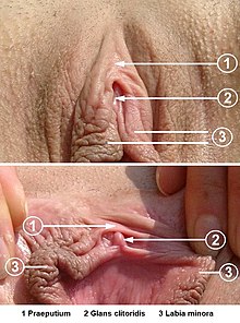
Clitoral hood (1) and clitoris (2). Labia are spread apart on the bottom image.
The clitoral hood projects at the front of the labia commissure, where the edges of the labia majora (outer lips) meet at the base of the pubic mound; it is partially formed by fusion of the upper part of the external folds of the labia minora (inner lips) and covers the glans and external shaft.[39] There is considerable variation in how much of the glans protrudes from the hood and how much is covered by it, ranging from completely covered to fully exposed,[37] and tissue of the labia minora also encircles the base of the glans.[40]
Bulbs
The vestibular bulbs are more closely related to the clitoris than the vestibule because of the similarity of the trabecular and erectile tissue within the clitoris and bulbs, and the absence of trabecular tissue in other genital organs, with the erectile tissue’s trabecular nature allowing engorgement and expansion during sexual arousal.[28][40] The vestibular bulbs are typically described as lying close to the crura on either side of the vaginal opening; internally, they are beneath the labia majora. When engorged with blood, they cuff the vaginal opening and cause the vulva to expand outward.[28] Although several texts state that they surround the vaginal opening, Ginger et al. state that this does not appear to be the case and tunica albuginea does not envelop the erectile tissue of the bulbs.[28] In Yang et al.’s assessment of the bulbs’ anatomy, they conclude that the bulbs “arch over the distal urethra, outlining what might be appropriately called the ‘bulbar urethra’ in women.”[29]
Homology
The clitoris and penis generally have the same anatomical structure, although the distal portion (or opening) of the urethra is absent in the clitoris of humans and most other animals. The idea that males have clitorises was suggested in 1987 by researcher Josephine Lowndes Sevely theorized that the male corpora cavernosa (a pair of sponge-like regions of erectile tissue that contain most of the blood in the penis during penile erection) are the true counterpart of the clitoris. She argued that “the male clitoris” is directly beneath the rim of the glans penis, where the frenulum of prepuce of the penis (a fold of the prepuce) is located, and proposed that this area be called the “Lownde’s crown”. Her theory and proposal, though acknowledged in anatomical literature, did not materialize in anatomy books.[41] Modern anatomical texts show that the clitoris displays a hood that is the equivalent of the penis’s foreskin, which covers the glans. It also has a shaft that is attached to the glans. The male corpora cavernosa are homologous to the corpus cavernosum clitoridis (the female cavernosa), the bulb of the penis is homologous to the vestibular bulbs beneath the labia minora, the scrotum is homologous to the labia majora, and the penile urethra and part of the skin of the penis are homologous to the labia minora.[42]
Upon anatomical study, the penis can be described as a clitoris that has been mostly pulled out of the body and grafted on top of a significantly smaller piece of spongiosum containing the urethra.[42] Concerning nerve endings, the human clitoris’s estimated number of nerve endings (8,000 to over 10,000) is commonly cited as being twice as many as the nerve endings found in the human penis (for its glans or body as a whole) and as more than any other part of the human body.[3][4] These reports sometimes conflict with other sources on clitoral anatomy or those concerning the nerve endings in the human penis. For example, while some sources estimate that the human penis has 4,000 nerve endings,[3] other sources state that the glans or the entire penile structure have the same amount of nerve endings as the clitoral glans.[43] Some sources state that in contrast to the glans penis, the clitoral glans lacks smooth muscle within its fibrovascular cap and is thus differentiated from the erectile tissues of the clitoris and bulbs; additionally, bulb size varies and may be dependent on age and estrogenization.[28] While the bulbs are considered the equivalent of the male spongiosum, they do not completely encircle the urethra.[28]
The thin corpus spongiosum of the penis runs along the underside of the penile shaft, enveloping the urethra, and expands at the end to form the glans. It partially contributes to erection, which is primarily caused by the two corpora cavernosa that comprise the bulk of the shaft; like the female cavernosa, the male cavernosa soak up blood and become erect when sexually excited.[44] The male corpora cavernosa taper off internally on reaching the spongiosum head.[44] Concerning the Y-shape of the cavernosa – crown, body, and legs – the body accounts for much more of the structure in men, and the legs are stubbier; typically, the cavernosa are longer and thicker in males than in females.[29][45]
Function
Sexual activity
General
The clitoris has an abundance of nerve endings, and is the human female’s most sensitive erogenous zone and generally the primary anatomical source of human female sexual pleasure.[2] When sexually stimulated, it may incite female sexual arousal. Sexual stimulation, including arousal, may result from mental stimulation, foreplay with a sexual partner, or masturbation, and can lead to orgasm.[46] The most effective sexual stimulation of the organ is usually manually or orally (cunnilingus), which is often referred to as direct clitoral stimulation; in cases involving sexual penetration, these activities may also be referred to as additional or assisted clitoral stimulation.[47]
Direct clitoral stimulation involves physical stimulation to the external anatomy of the clitoris – glans, hood, and the external shaft.[48] Stimulation of the labia minora (inner lips), due to its external connection with the glans and hood, may have the same effect as direct clitoral stimulation.[49] Though these areas may also receive indirect physical stimulation during sexual activity, such as when in friction with the labia majora (outer lips),[50] indirect clitoral stimulation is more commonly attributed to penile-vaginal penetration.[51][52] Penile-anal penetration may also indirectly stimulate the clitoris by the shared sensory nerves (especially the pudendal nerve, which gives off the inferior anal nerves and divides into two terminal branches: the perineal nerve and the dorsal nerve of the clitoris).[53]
Due to the glans’ high sensitivity, direct stimulation to it is not always pleasurable; instead, direct stimulation to the hood or the areas near the glans is often more pleasurable, with the majority of women preferring to use the hood to stimulate the glans, or to have the glans rolled between the lips of the labia, for indirect touch.[54] It is also common for women to enjoy the shaft of the clitoris being softly caressed in concert with the occasional circling of the clitoral glans. This might be with or without manual penetration of the vagina, while other women enjoy having the entire area of the vulva caressed.[55] As opposed to the use of dry fingers, stimulation from well-lubricated fingers, either by vaginal lubrication or a personal lubricant, is usually more pleasurable for the external anatomy of the clitoris.[56][57]
As the clitoris’s external location does not allow for direct stimulation by sexual penetration, any external clitoral stimulation while in the missionary position usually results from the pubic bone area, the movement of the groins when in contact. As such, some couples may engage in the woman-on-top position or the coital alignment technique, a sex position combining the “riding high” variation of the missionary position with pressure-counterpressure movements performed by each partner in rhythm with sexual penetration, to maximize clitoral stimulation.[58][59] Lesbian couples may engage in tribadism for ample clitoral stimulation or mutual clitoral stimulation during whole-body contact.[N 2][61][62] Pressing the penis in a gliding or circular motion against the clitoris (intercrural sex), or stimulating it by the movement against another body part, may also be practiced.[63][64] A vibrator (such as a clitoral vibrator), dildo or other sex toy may be used.[63][65] Other women stimulate the clitoris by use of a pillow or other inanimate object, by a jet of water from the faucet of a bathtub or shower, or by closing their legs and rocking.[66][67][68]
During sexual arousal, the clitoris and the whole of the genitalia engorge and change color as the erectile tissues fill with blood (vasocongestion), and the individual experiences vaginal contractions.[69] The ischiocavernosus and bulbocavernosus muscles, which insert into the corpora cavernosa, contract and compress the dorsal vein of the clitoris (the only vein that drains the blood from the spaces in the corpora cavernosa), and the arterial blood continues a steady flow and having no way to drain out, fills the venous spaces until they become turgid and engorged with blood. This is what leads to clitoral erection.[12][70]
The clitoral glans doubles in diameter upon arousal and further stimulation become less visible as it is covered by the swelling of tissues of the clitoral hood.[69][71] The swelling protects the glans from direct contact, as direct contact at this stage can be more irritating than pleasurable.[71][72] Vasocongestion eventually triggers a muscular reflex, which expels the blood that was trapped in surrounding tissues, and leads to an orgasm.[73] A short time after stimulation has stopped, especially if orgasm has been achieved, the glans becomes visible again and returns to its normal state,[74] with a few seconds (usually 5–10) to return to its normal position and 5–10 minutes to return to its original size.[N 3][71][76] If orgasm is not achieved, the clitoris may remain engorged for a few hours, which women often find uncomfortable.[58] Additionally, the clitoris is very sensitive after orgasm, making further stimulation initially painful for some women.[77]
Clitoral and vaginal orgasmic factors
General statistics indicate that 70–80 percent of women require direct clitoral stimulation (consistent manual, oral, or other concentrated friction against the external parts of the clitoris) to reach orgasm.[N 4][N 5][N 6][81] Indirect clitoral stimulation (for example, via vaginal penetration) may also be sufficient for female orgasm.[N 7][16][83] The area near the entrance of the vagina (the lower third) contains nearly 90 percent of the vaginal nerve endings, and there are areas in the anterior vaginal wall and between the top junction of the labia minora and the urethra that are especially sensitive, but intense sexual pleasure, including orgasm, solely from vaginal stimulation is occasional or otherwise absent because the vagina has significantly fewer nerve endings than the clitoris.[84]
The prominent debate over the quantity of vaginal nerve endings began with Alfred Kinsey. Although Sigmund Freud’s theory that clitoral orgasms are a prepubertal or adolescent phenomenon and that vaginal (or G-spot) orgasms are something that only physically mature females experience had been criticized before, Kinsey was the first researcher to harshly criticize the theory.[85][86] Through his observations of female masturbation and interviews with thousands of women,[87] Kinsey found that most of the women he observed and surveyed could not have vaginal orgasms,[88] a finding that was also supported by his knowledge of sex organ anatomy.[89] Scholar Janice M. Irvine stated that he “criticized Freud and other theorists for projecting male constructs of sexuality onto women” and “viewed the clitoris as the main center of sexual response”. He considered the vagina to be “relatively unimportant” for sexual satisfaction, relaying that “few women inserted fingers or objects into their vaginas when they masturbated”. Believing that vaginal orgasms are “a physiological impossibility” because the vagina has insufficient nerve endings for sexual pleasure or climax, he “concluded that satisfaction from penile penetration [is] mainly psychological or perhaps the result of referred sensation”.[90]
Masters and Johnson’s research, as well as Shere Hite’s, generally supported Kinsey’s findings about the female orgasm.[91] Masters and Johnson were the first researchers to determine that the clitoral structures surround and extend along and within the labia. They observed that both clitoral and vaginal orgasms have the same stages of physical response, and found that the majority of their subjects could only achieve clitoral orgasms, while a minority achieved vaginal orgasms. On that basis, they argued that clitoral stimulation is the source of both kinds of orgasms,[92] reasoning that the clitoris is stimulated during penetration by friction against its hood.[93] The research came at the time of the second-wave feminist movement, which inspired feminists to reject the distinction made between clitoral and vaginal orgasms.[85][94] Feminist Anne Koedt argued that because men “have orgasms essentially by friction with the vagina” and not the clitoral area, this is why women’s biology had not been properly analyzed. “Today, with extensive knowledge of anatomy, with [C. Lombard Kelly], Kinsey, and Masters and Johnson, to mention just a few sources, there is no ignorance on the subject [of the female orgasm],” she stated in her 1970 article The Myth of the Vaginal Orgasm. She added, “There are, however, social reasons why this knowledge has not been popularized. We are living in a male society which has not sought change in women’s role.”[85]
Supporting an anatomical relationship between the clitoris and vagina is a study published in 2005, which investigated the size of the clitoris; Australian urologist Helen O’Connell, described as having initiated discourse among mainstream medical professionals to refocus on and redefine the clitoris, noted a direct relationship between the legs or roots of the clitoris and the erectile tissue of the clitoral bulbs and corpora, and the distal urethra and vagina while using magnetic resonance imaging (MRI) technology.[95][96] While some studies, using ultrasound, have found physiological evidence of the G-spot in women who report having orgasms during vaginal intercourse,[83] O’Connell argues that this interconnected relationship is the physiological explanation for the conjectured G-Spot and experience of vaginal orgasms, taking into account the stimulation of the internal parts of the clitoris during vaginal penetration. “The vaginal wall is, in fact, the clitoris,” she said. “If you lift the skin off the vagina on the side walls, you get the bulbs of the clitoris – triangular, crescental masses of erectile tissue.”[16] O’Connell et al., having performed dissections on the female genitals of cadavers and used photography to map the structure of nerves in the clitoris, made the assertion in 1998 that there is more erectile tissue associated with the clitoris than is generally described in anatomical textbooks and were thus already aware that the clitoris is more than just its glans.[97] They concluded that some females have more extensive clitoral tissues and nerves than others, especially having observed this in young cadavers compared to elderly ones,[97] and therefore whereas the majority of females can only achieve orgasm by direct stimulation of the external parts of the clitoris, the stimulation of the more generalized tissues of the clitoris via vaginal intercourse may be sufficient for others.[16]
French researchers Odile Buisson Fr and Pierre Foldès reported similar findings to that of O’Connell’s. In 2008, they published the first complete 3D sonography of the stimulated clitoris and republished it in 2009 with new research, demonstrating how erectile tissue of the clitoris engorges and surrounds the vagina. Based on their findings, they argued that women may be able to achieve vaginal orgasm via stimulation of the G-spot because the highly innervated clitoris is pulled closely to the anterior wall of the vagina when the woman is sexually aroused and during vaginal penetration. They assert that since the front wall of the vagina is inextricably linked with the internal parts of the clitoris, stimulating the vagina without activating the clitoris may be next to impossible. In their 2009 published study, the “coronal planes during perineal contraction and finger penetration demonstrated a close relationship between the root of the clitoris and the anterior vaginal wall”. Buisson and Foldès suggested “that the special sensitivity of the lower anterior vaginal wall could be explained by pressure and movement of clitoris’s root during a vaginal penetration and subsequent perineal contraction”.[98][99]
Researcher Vincenzo Puppo, who, while agreeing that the clitoris is the center of female sexual pleasure and believing that there is no anatomical evidence of the vaginal orgasm, disagrees with O’Connell and other researchers’ terminological and anatomical descriptions of the clitoris (such as referring to the vestibular bulbs as the “clitoral bulbs”) and states that “the inner clitoris” does not exist because the penis cannot come in contact with the congregation of multiple nerves/veins situated until the angle of the clitoris, detailed by Kobelt, or with the roots of the clitoris, which do not have sensory receptors or erogenous sensitivity, during vaginal intercourse.[15] Puppo’s belief contrasts the general belief among researchers that vaginal orgasms are the result of clitoral stimulation; they reaffirm that clitoral tissue extends, or is at least stimulated by its bulbs, even in the area most commonly reported to be the G-spot.[100]
The G-spot is analogous to the base of the male penis and has additionally been theorized, with the sentiment from researcher Amichai Kilchevsky that because female fetal development is the “default” state in the absence of substantial exposure to male hormones and therefore the penis is essentially a clitoris enlarged by such hormones, there is no evolutionary reason why females would have an entity in addition to the clitoris that can produce orgasms.[101] The general difficulty of achieving orgasms vaginally, which is a predicament that is likely due to nature easing the process of childbearing by drastically reducing the number of vaginal nerve endings,[102] challenge arguments that vaginal orgasms help encourage sexual intercourse to facilitate reproduction.[103][104] Supporting a distinct G-spot, however, is a study by Rutgers University, published in 2011, which was the first to map the female genitals onto the sensory portion of the brain; the scans indicated that the brain registered distinct feelings between stimulating the clitoris, the cervix and the vaginal wall – where the G-spot is reported to be – when several women stimulated themselves in a functional magnetic resonance (fMRI) machine.[99][105] Barry Komisaruk, head of the research findings, stated that he feels that “the bulk of the evidence shows that the G-spot is not a particular thing” and that it is “a region, it’s a convergence of many different structures”.[103]
Vestigiality, adaptionist and reproductive views
Whether the clitoris is vestigial, an adaptation, or serves a reproductive function has also been debated.[106][107] Geoffrey Miller stated that Helen Fisher, Meredith Small and Sarah Blaffer Hrdy “have viewed the clitoral orgasm as a legitimate adaptation in its own right, with major implications for female sexual behavior and sexual evolution”.[108] Like Lynn Margulis and Natalie Angier, Miller believes, “The human clitoris shows no apparent signs of having evolved directly through male mate choice. It is not especially large, brightly colored, specifically shaped or selectively displayed during courtship.” He contrasts this with other female species such as spider monkeys and spotted hyenas that have clitorises as long as their male counterparts. He said the human clitoris “could have evolved to be much more conspicuous if males had preferred sexual partners with larger brighter clitorises” and that “its inconspicuous design combined with its exquisite sensitivity suggests that the clitoris is important not as an object of male mate choice, but as a mechanism of female choice.”[108]
While Miller stated that male scientists such as Stephen Jay Gould and Donald Symons “have viewed the female clitoral orgasm as an evolutionary side-effect of the male capacity for penile orgasm” and that they “suggested that clitoral orgasm cannot be an adaptation because it is too hard to achieve”,[108] Gould acknowledged that “most female orgasms emanate from a clitoral, rather than vaginal (or some other), site” and that his nonadaptive belief “has been widely misunderstood as a denial of either the adaptive value of female orgasm in general or even as a claim that female orgasms lack significance in some broader sense”. He said that although he accepts that “clitoral orgasm plays a pleasurable and central role in female sexuality and its joys,” “[a]ll these favorable attributes, however, emerge just as clearly and just as easily, whether the clitoral site of orgasm arose as a spandrel or an adaptation”. He added that the “male biologists who fretted over [the adaptionist questions] simply assumed that a deeply vaginal site, nearer the region of fertilization, would offer greater selective benefit” due to their Darwinian, summum bonum beliefs about enhanced reproductive success.[109]
Similar to Gould’s beliefs about adaptionist views and that “females grow nipples as adaptations for suckling, and males grow smaller unused nipples as a spandrel based upon the value of single development channels”,[109] Elisabeth Lloyd suggested that there is little evidence to support an adaptionist account of female orgasm.[104][107] Meredith L. Chivers stated that “Lloyd views female orgasm as an ontogenetic leftover; women have orgasms because the urogenital neurophysiology for orgasm is so strongly selected for in males that this developmental blueprint gets expressed in females without affecting fitness” and this is similar to “males hav[ing] nipples that serve no fitness-related function.”[107]
At the 2002 conference for Canadian Society of Women in Philosophy, Nancy Tuana argued that the clitoris is unnecessary in reproduction; she stated that it has been ignored because of “a fear of pleasure. It is pleasure separated from reproduction. That’s the fear.” She reasoned that this fear causes ignorance, which veils female sexuality.[110] O’Connell stated, “It boils down to rivalry between the sexes: the idea that one sex is sexual and the other reproductive. The truth is that both are sexual and both are reproductive.” She reiterated that the vestibular bulbs appear to be part of the clitoris and that the distal urethra and vagina are intimately related structures, although they are not erectile in character, forming a tissue cluster with the clitoris that appears to be the location of female sexual function and orgasm.[16][29]
Clinical significance
Modification

Modifications to the clitoris can be intentional or unintentional. They include female genital mutilation (FGM), sex reassignment surgery (for trans men as part transitioning, which may also include clitoris enlargement), intersex surgery, and genital piercings.[27][111][112] Use of anabolic steroids by bodybuilders and other athletes can result in significant enlargement of the clitoris in concert with other masculinizing effects on their bodies.[113][114] Abnormal enlargement of the clitoris may also be referred to as clitoromegaly, but clitoromegaly is more commonly seen as a congenital anomaly of the genitalia.[23]
People taking hormones or other medications as part of a transgender transition usually experience dramatic clitoral growth; individual desires and the difficulties of phalloplasty (construction of a penis) often result in the retention of the original genitalia with the enlarged clitoris as a penis analog (metoidioplasty).[27][112] However, the clitoris cannot reach the size of the penis through hormones.[112] A surgery to add function to the clitoris, such as metoidioplasty, is an alternative to phalloplasty that permits the retention of sexual sensation in the clitoris.[112]
In clitoridectomy, the clitoris may be removed as part of a radical vulvectomy to treat cancer such as vulvar intraepithelial neoplasia; however, modern treatments favor more conservative approaches, as invasive surgery can have psychosexual consequences.[115] Clitoridectomy more often involves parts of the clitoris being partially or completely removed during FGM, which may be additionally known as female circumcision or female genital cutting (FGC).[116][117] Removing the glans of the clitoris does not mean that the whole structure is lost, since the clitoris reaches deep into the genitals.[16]
In reduction clitoroplasty, a common intersex surgery, the glans are preserved and parts of the erectile bodies are excised.[27] Problems with this technique include loss of sensation, loss of sexual function, and sloughing of the glans.[27] One way to preserve the clitoris with its innervations and function is to imbricate and bury the clitoral glans; however, Şenaylı et al. state that “pain during stimulus because of trapped tissue under the scarring is nearly routine. In another method, 50 percent of the ventral clitoris is removed through the level base of the clitoral shaft, and it is reported that good sensation and clitoral function are observed in follow-up”; additionally, it has “been reported that the complications are from the same as those in the older procedures for this method”.[27]
Concerning females who have the condition congenital adrenal hyperplasia, the largest group requiring surgical genital correction, researcher Atilla Şenaylı stated, “The main expectations for the operations are to create a normal female anatomy, with minimal complications and improvement of life quality.” Şenaylı added that “[c]osmesis, structural integrity, the coital capacity of the vagina, and absence of pain during sexual activity are the parameters to be judged by the surgeon.” (Cosmesis usually refers to the surgical correction of a disfiguring defect.) He stated that although “expectations can be standardized within these few parameters, operative techniques have not yet become homogeneous. Investigators have preferred different operations for different ages of patients”.[27]
Gender assessment and surgical treatment are the two main steps in intersex operations. “The first treatments for clitoromegaly were simply resection of the clitoris. Later, it was understood that the clitoris glans and sensory input are important to facilitate orgasm,” stated Atilla. The clitoral glans’ epithelium “has high cutaneous sensitivity, which is important in sexual responses”, and it is because of this that “recession clitoroplasty was later devised as an alternative, but reduction clitoroplasty is the method currently performed.”[27]
What is often referred to as “clit piercing” is the more common (and significantly less complicated) clitoral hood piercing. Since clitoral piercing is difficult and very painful, piercing the clitoral hood is more common than piercing the clitoral shaft, owing to the small percentage of people who are anatomically suited for it.[111] Clitoral hood piercings are usually channeled in the form of vertical piercings, and, to a lesser extent, horizontal piercings. The triangle piercing is a very deep horizontal hood piercing and is done behind the clitoris as opposed to in front of it. For styles such as the Isabella piercing, which pass through the clitoral shaft but are placed deep at the base, they provide unique stimulation and still require the proper genital build. The Isabella starts between the clitoral glans and the urethra, exiting at the top of the clitoral hood; this piercing is highly risky concerning the damage that may occur because of intersecting nerves.[111]
Sexual disorders
Persistent genital arousal disorder (PGAD) results in spontaneous, persistent, and uncontrollable genital arousal in women, unrelated to any feelings of sexual desire.[118] Clitoral priapism, also known as clitorism, is a rare, potentially painful medical condition and is sometimes described as an aspect of PGAD.[118] With PGAD, arousal lasts for an unusually extended period (ranging from hours to days);[119] it can also be associated with morphometric and vascular modifications of the clitoris.[120]
Drugs may cause or affect clitoral priapism. The drug trazodone is known to cause male priapism as a side effect, but there is only one documented report that it may have caused clitoral priapism, in which case discontinuing the medication may be a remedy.[121] Additionally, nefazodone is documented to have caused clitoral engorgement, as distinct from clitoral priapism, in one case,[121] and clitoral priapism can sometimes start as a result of, or only after, the discontinuation of antipsychotics or selective serotonin reuptake inhibitors (SSRIs).[122]
Because PGAD is relatively rare and, as its concept apart from clitoral priapism, has only been researched since 2001, there is little research into what may cure or remedy the disorder.[118] In some recorded cases, PGAD was caused by or caused, a pelvic arterial-venous malformation with arterial branches to the clitoris; surgical treatment was effective in these cases.[123]
In 2022, an article in The New York Times reported several instances of women experiencing reduced clitoral sensitivity or inability to orgasm following various surgical procedures, including biopsies of the vulva, pelvic mesh surgeries (sling surgeries), and labiaplasties. The Times quoted several researchers who suggest that surgeons’ lack of training in clitoral anatomy and nerve distribution may have been a factor.[124]
Society and culture
Ancient Greek–16th century knowledge and vernacular
Concerning historical and modern perceptions of the clitoris, the clitoris, and the penis was considered equivalent by some scholars for more than 2,500 years in all respects except their arrangement.[125] Due to it being frequently omitted from, or misrepresented in, historical and contemporary anatomical texts, it was also subject to a continual cycle of male scholars claiming to have discovered it.[126] The ancient Greeks, ancient Romans, and Greek and Roman generations up to and throughout the Renaissance, were aware that male and female sex organs are anatomically similar,[127][128] but prominent anatomists such as Galen (129 – c. 200 AD) and Vesalius (1514–1564) regarded the vagina as the structural equivalent of the penis, except for being inverted; Vesalius argued against the existence of the clitoris in normal women, and his anatomical model described how the penis corresponds with the vagina, without a role for the clitoris.[129]
Ancient Greek and Roman sexuality additionally designated penetration as “male-defined” sexuality. The term tribas, or tribade, was used to refer to a woman or intersex individual who actively penetrated another person (male or female) through the use of the clitoris or a dildo. As any sexual act was believed to require that one of the partners be “phallic” and that therefore sexual activity between women was impossible without this feature, mythology popularly associated lesbians with either having enlarged clitorises or as incapable of enjoying sexual activity without the substitution of a phallus.[130][131]

In 1545, Charles Estienne was the first writer to identify the clitoris in a work based on dissection, but he concluded that it had a urinary function.[16] Following this study, Realdo Colombo (also known as Matteo Renaldo Colombo), a lecturer in surgery at the University of Padua, Italy, published a book called De re anatomica in 1559, in which he describes the “seat of woman’s delight”.[132] In his role as researcher, Colombo concluded, “Since no one has discerned these projections and their workings, if it is permissible to give names to things discovered by me, it should be called the love or sweetness of Venus.”, about the mythological Venus, goddess of erotic love.[133][134] Colombo’s claim was disputed by his successor at Padua, Gabriele Falloppio (discoverer of the fallopian tube), who claimed that he was the first to discover the clitoris. In 1561, Falloppio stated, “Modern anatomists have entirely neglected it … and do not say a word about it … and if others have spoken of it, know that they have taken it from me or my students.” This caused an upset in the European medical community, and, having read Colombo’s and Falloppio’s detailed descriptions of the clitoris, Vesalius stated, “It is unreasonable to blame others for incompetence on the basis of some sport of nature you have observed in some women and you can hardly ascribe this new and useless part, as if it were an organ, to healthy women.” He concluded, “I think that such a structure appears in hermaphrodites who otherwise have well-formed genitals, as Paul of Aegina describes, but I have never once seen in any woman a penis (which Avicenna called albaratha and the Greeks called an enlarged nympha and classed as an illness) or even the rudiments of a tiny phallus.”[135]
The average anatomist had difficulty challenging Galen’s or Vesalius’s research; Galen was the most famous physician of the Greek era and his works were considered the standard of medical understanding up to and throughout the Renaissance (i.e. for almost two thousand years),[128][129] and various terms being used to describe the clitoris seemed to have further confused the issue of its structure. In addition to Avicenna’s naming it the albaratha or virga (“rod”) and Colombo’s calling it the sweetness of Venus, Hippocrates used the term columella (“little pillar'”), and Albucasis, an Arabic medical authority, named it tentigo (“tension”). The names indicated that each description of the structures was about the body and glans of the clitoris but usually the glans.[16] It was additionally known to the Romans, who named it (vulgar slang) landica.[136] However, Albertus Magnus, one of the most prolific writers of the Middle Ages, felt that it was important to highlight “homologies between male and female structures and function” by adding “a psychology of sexual arousal” that Aristotle had not used to detail the clitoris. While in Constantine’s treatise Liber de coitu, the clitoris is referred to a few times, Magnus gave an equal amount of attention to male and female organs.[16]
Like Avicenna, Magnus also used the word virga for the clitoris, but employed it for the male and female genitals; despite his efforts to give equal ground to the clitoris, the cycle of suppression and rediscovery of the organ continued, and a 16th-century justification for clitoridectomy appears to have been confused by hermaphroditism and the imprecision created by the word nymphae substituted for the word clitoris. Nymphotomia was a medical operation to excise an unusually large clitoris, but what was considered “unusually large” was often a matter of perception.[16] The procedure was routinely performed on Egyptian women,[137][138] due to physicians such as Jacques Daléchamps who believed that this version of the clitoris was “an unusual feature that occurred in almost all Egyptian women [and] some of ours, so that when they find themselves in the company of other women, or their clothes rub them while they walk or their husbands wish to approach them, it erects like a male penis and indeed they use it to play with other women, as their husbands would do … Thus the parts are cut”.[16]
17th century–present day knowledge and vernacular

Caspar Bartholin, a 17th-century Danish anatomist, dismissed Colombo’s and Falloppio’s claims that they discovered the clitoris, arguing that the clitoris had been widely known to medical science since the second century.[139] Although 17th-century midwives recommended to men and women that women should aspire to achieve orgasms to help them get pregnant for general health and well-being and to keep their relationships healthy,[128] debate about the importance of the clitoris persisted, notably in the work of Regnier de Graaf in the 17th century[40][140] and Georg Ludwig Kobelt in the 19th.[16]
Like Falloppio and Bartholin, de Graaf criticized Colombo’s claim of having discovered the clitoris; his work appears to have provided the first comprehensive account of clitoral anatomy.[141] “We are extremely surprised that some anatomists make no more mention of this part than if it did not exist at all in the universe of nature,” he stated. “In every cadaver, we have so far dissected we have found it quite perceptible to sight and touch.” De Graaf stressed the need to distinguish nympha from clitoris, choosing to “always give [the clitoris] the name clitoris” to avoid confusion; this resulted in the frequent use of the correct name for the organ among anatomists, but considering that nympha was also varied in its use and eventually became the term specific to the labia minora, more confusion ensued.[16] Debate about whether orgasm was even necessary for women began in the Victorian era, and Freud’s 1905 theory about the immaturity of clitoral orgasms (see above) negatively affected women’s sexuality throughout most of the 20th century.[128][142]
Toward the end of World War I, a maverick British MP named Noel Pemberton Billing published an article entitled “The Cult of the Clitoris”, furthering his conspiracy theories and attacking the actress Maud Allan and Margot Asquith, wife of the prime minister. The accusations led to a sensational libel trial, which Billing eventually won; Philip Hoare reports that Billing argued that “as a medical term, ‘clitoris’ would only be known to the ‘initiated’, and was incapable of corrupting moral minds”.[143] Jodie Medd argues regarding “The Cult of the Clitoris” that “the female non-reproductive but desiring body […] simultaneously demands and refuses interpretative attention, inciting scandal through its very resistance to representation.”[144]
From the 18th 20th century, especially during the 20th, details of the clitoris from various genital diagrams presented in earlier centuries were omitted from later texts.[128][145] The full extent of the clitoris was alluded to by Masters and Johnson in 1966, but in such a muddled fashion that the significance of their description became obscured; in 1981, the Federation of Feminist Women’s Health Clinics (FFWHC) continued this process with anatomically precise illustrations identifying 18 structures of the clitoris.[55][128] Despite the FFWHC’s illustrations, Josephine Lowndes Sevely, in 1987, described the vagina as more of the counterpart of the penis.[146]
Concerning other beliefs about the clitoris, Hite (1976 and 1981) found that, during sexual intimacy with a partner, clitoral stimulation was more often described by women as foreplay than as a primary method of sexual activity, including orgasm.[147] Further, although the FFWHC’s work significantly propelled feminist reformation of anatomical texts, it did not have a general impact.[96][148] Helen O’Connell’s late 1990s research motivated the medical community to start changing the way the clitoris is anatomically defined.[96] O’Connell describes typical textbook descriptions of the clitoris as lacking detail and including inaccuracies, such as older and modern anatomical descriptions of the female human urethral and genital anatomy having been based on dissections performed on elderly cadavers whose erectile (clitoral) tissue had shrunk.[97] She instead credits the work of Georg Ludwig Kobelt as the most comprehensive and accurate description of clitoral anatomy.[16] MRI measurements, which provide a live and multi-planar method of examination, now complement the FFWHC’s, as well as O’Connell’s, research efforts concerning the clitoris, showing that the volume of clitoral erectile tissue is ten times that which is shown in doctors’ offices and anatomy textbooks.[40][96]
In Bruce Bagemihl’s survey of The Zoological Record (1978–1997) – which contains over a million documents from over 6,000 scientific journals – 539 articles focusing on the penis were found, while seven were found focusing on the clitoris.[149] In 2000, researchers Shirley Ogletree and Harvey Ginsberg concluded that there is a general neglect of the word clitoris in the common vernacular. They looked at the terms used to describe genitalia in the PsycINFO database from 1887 to 2000 and found that penis was used in 1,482 sources, vagina in 409, while clitoris was only mentioned in 83. They additionally analyzed 57 books listed in a computer database for sex instruction. In the majority of the books, penis was the most commonly discussed body part – mentioned more than clitoris, vagina, and uterus put together. They last investigated terminology used by college students, ranging from Euro-American (76%/76%), Hispanic (18%/14%), and African American (4%/7%), regarding the students’ beliefs about sexuality and knowledge on the subject. The students were overwhelmingly educated to believe that the vagina is the female counterpart of the penis. The authors found that the student’s belief that the inner portion of the vagina is the most sexually sensitive part of the female body correlated with negative attitudes toward masturbation and strong support for sexual myths.[150][151]

Girl protesting for clitoris-awareness at a women’s rights rally in Paris, 2019
A study in 2005 reported that, among a sample of undergraduate students, the most frequently cited sources for knowledge about the clitoris were school and friends, and that this was associated with the least tested knowledge. Knowledge of the clitoris by self-exploration was the least cited, but “respondents correctly answered, on average, three of the five clitoral knowledge measures”. The authors stated that “[k]nowledge correlated significantly with the frequency of women’s orgasm in masturbation but not partnered sex” and that their “results are discussed in light of gender inequality and a social construction of sexuality, endorsed by both men and women, that privileges men’s sexual pleasure over women’s, such that orgasm for women is pleasing but ultimately incidental.” They concluded that part of the solution to remedying “this problem” requires that males and females are taught more about the clitoris than is currently practiced.[152]
The humanitarian group Clitoraid launched the first annual International Clitoris Awareness Week, from 6 to 12 May in 2015. Clitoraid spokesperson Nadine Gary stated that the group’s mission is to raise public awareness about the clitoris because it has “been ignored, vilified, made taboo, and considered sinful and shameful for centuries”.[153][154]
Odile Fillod created a 3D printable, open source, full-size model of the clitoris, for use in a set of anti-sexist videos she had been commissioned to produce. Fillod was interviewed by Stephanie Theobald, whose article in The Guardian stated that the 3D model would be used for sex education in French schools, from primary to secondary level, from September 2016 onwards;[155] this was not the case, but the story went viral across the world.[156]
A questionnaire in a 2019 study was administered to a sample of educational sciences postgraduate students to trace the level of their knowledge concerning the organs of the female and male reproductive system. The authors reported that about two-thirds of the students failed to name external female genitals, such as the clitoris and labia, even after detailed pictures were provided to them.[157] An analysis in 2022 reported that the clitoris is mentioned in only one out of 113 Greek secondary education textbooks used in biology classes from the 1870s to present.[158]
Contemporary art
New York artist Sophia Wallace started work in 2012 on a multimedia project to challenge misconceptions about the clitoris. Based on O’Connell’s 1998 research, Wallace’s work emphasizes the sheer scope and size of the human clitoris. She says that ignorance of this still seems to be pervasive in modern society. “It is a curious dilemma to observe the paradox that on the one hand, the female body is the primary metaphor for sexuality, its use saturates advertising, art, and the mainstream erotic imaginary,” she said. “Yet, the clitoris, the true female sexual organ, is virtually invisible.” The project is called Cliteracy and it includes a “clit rodeo”, which is interactive, climb-on model of a giant golden clitoris, including its inner parts, produced with the help of sculptor Kenneth Thomas. “It’s been a showstopper wherever it’s been shown. People are hungry to be able to talk about this,” Wallace said. “I love seeing men standing up for the clit […] Cliteracy is about not having one’s body controlled or legislated […] Not having access to the pleasure that is your birthright is a deeply political act.”[159]
Another project started in New York, in 2016, street art that has since spread to almost 100 cities: Clitorosity, a “community-driven effort to celebrate the full structure of the clitoris”, combining chalk drawings and words to spark interaction and conversation with passers-by, which the team documents on social media.[160][161] In 2016, Lori-Malépart Traversy made an animated documentary about the unrecognized anatomy of the clitoris.[162]
Alli Sebastian Wolf created a golden 100∶1 scale model anatomical of a clitoris in 2017, called the Glitoris and said, she hopes knowledge of the clitoris will soon become so uncontroversial that making art about them would be as irrelevant as making art about penises.[163]
Other projects listed by the BBC include Clito Clito, body-positive jewellery made in Berlin; Clitorissima, a documentary intended to normalize mother-daughter conversations about the clitoris; and a ClitArt festival in London, encompassing spoken word performances as well as visual art.[161] French art collective Les Infemmes (a pun on “infamous” and “women”) published a fanzine whose title can be translated as “The Clit Cheatsheet”.[164]
Influence on female genital mutilation
Significant controversy surrounds female genital mutilation (FGM),[116][117] with the World Health Organization (WHO) being one of many health organizations that have campaigned against the procedures on behalf of human rights, stating that “FGM has no health benefits” and that it is “a violation of the human rights of girls and women” which “reflects deep-rooted inequality between the sexes”.[117] The practice has existed at one point or another in almost all human civilizations,[137] most commonly to exert control over the sexual behavior, including masturbation, of girls and women, but also to change the clitoris’s appearance.[117][138][165] Custom and tradition are the most frequently cited reasons for FGM, with some cultures believing that not performing it has the possibility of disrupting the cohesiveness of their social and political systems, such as FGM also being a part of a girl’s initiation into adulthood. Often, a girl is not considered an adult in an FGM-practicing society unless she has undergone FGM,[117][138] and the “removal of the clitoris and labia – viewed by some as the male parts of a woman’s body – is thought to enhance the girl’s femininity, often synonymous with docility and obedience”.[138]
Female genital mutilation is carried out in several societies, especially in Africa, with 85 percent of genital mutilations performed in Africa consisting of clitoridectomy or excision,[138][166] and to a lesser extent in other parts of the Middle East and Southeast Asia, on girls from a few days old to mid-adolescent, often to reduce the sexual desire to preserve vaginal virginity.[117][138][165] The practice of FGM has spread globally, as immigrants from Asia, Africa, and the Middle East bring the custom with them.[167] In the United States, it is sometimes practiced on girls born with a clitoris that is larger than usual.[116] Comfort Momoh, who specializes in the topic of FGM, states that FGM might have been “practiced in ancient Egypt as a sign of distinction among the aristocracy”; there are reports that traces of infibulation are on Egyptian mummies.[137] FGM is still routinely practiced in Egypt.[138][168] Greenberg et al. report that “one study found that 97 percent of married women in Egypt had had some form of genital mutilation performed.”[168] Amnesty International estimated in 1997 that more than two million FGM procedures are performed every year.[138]
Other animals
General
Although the clitoris exists in all mammal species,[149] few detailed studies of the anatomy of the clitoris in non-humans exist.[169] The clitoris is especially developed in fossas,[170] apes, lemurs, moles,[171] and, like the penis in many non-human placental mammals, often contains a small bone. In females, this bone is known as the os clitoridis.[172] The clitoris exists in turtles,[173] ostriches,[174] crocodiles,[173] and in species of birds in which the male counterpart has a penis.[173] Some intersex female bears mate and give birth through the tip of the clitoris; these species are grizzly bears, brown bears, American black bears and polar bears. Although the bears have been described as having “a birth canal that runs through the clitoris rather than forming a separate vagina” (a feature that is estimated to make up 10 to 20 percent of the bears’ population),[175] scientists state that female spotted hyenas are the only non-hermaphroditic female mammals devoid of an external vaginal opening, and whose sexual anatomy is distinct from usual intersex cases.[176]
Non-human primates
In spider monkeys, the clitoris is especially developed and has an interior passage, or urethra, that makes it almost identical to the penis, and it retains and distributes urine droplets as the female spider monkey moves around. Scholar Alan F. Dixson stated that this urine “is voided at the bases of the clitoris, flows down the shallow groove on its perineal surface, and is held by the skin folds on each side of the groove”.[177] Because spider monkeys of South America have pendulous and erectile clitorises long enough to be mistaken for a penis, researchers and observers of the species look for a scrotum to determine the animal’s sex; a similar approach is to identify scent-marking glands that may also be present on the clitoris.[178]
The clitoris erects in squirrel monkeys during dominance displays, which indirectly influences the squirrel monkeys’ reproductive success.[179]
The clitoris of bonobos is larger and more externalized than in most mammals;[180] Natalie Angier said that a young adolescent “female bonobo is maybe half the weight of a human teenager, but her clitoris is three times bigger than the human equivalent, and visible enough to waggle unmistakably as she walks”.[181] Female bonobos often engage in the practice of genital-genital (GG) rubbing, which is the non-human form of tribadism that human females engage in. Ethologist Jonathan Balcombe stated that female bonobos rub their clitorises together rapidly for ten to twenty seconds, and this behavior, “which may be repeated in rapid succession, is usually accompanied by grinding, shrieking, and clitoral engorgement”; he added that, on average, they engage in this practice “about once every two hours”, and as bonobos sometimes mate face-to-face, “evolutionary biologist Marlene Zuk has suggested that the position of the clitoris in bonobos and some other primates has evolved to maximize stimulation during sexual intercourse”.[180]
Many strepsirrhine species exhibit elongated clitorises that are either fully or partially tunneled by the urethra, including mouse lemurs, dwarf lemurs, all Eulemur species, lorises and galagos.[182][183][184] Some of these species also exhibit a membrane seal across the vagina that closes the vaginal opening during the non-mating seasons, most notably mouse and dwarf lemurs.[182] The clitoral morphology of the ring-tailed lemur is the most well-studied. They are described as having “elongated, pendulous clitorises that are [fully] tunneled by a urethra”. The urethra is surrounded by erectile tissue, which allows for significant swelling during breeding seasons, but this erectile tissue differs from the typical male corpus spongiosum.[185] Non-pregnant adult ring-tailed females do not show higher testosterone levels than males, but they do exhibit higher A4 and estrogen levels during seasonal aggression. During pregnancy, estrogen, A4, and testosterone levels are raised, but female fetuses are still “protected” from excess testosterone.[186] These “masculinized” genitalia are often found alongside other traits, such as female-dominated social groups, reduced sexual dimorphism that makes females the same size as males, and even ratios of sexes in adult populations.[186][187] This phenomenon that has been dubbed the “lemur syndrome”.[188] A 2014 study of Eulemur masculinization proposed that behavioral and morphological masculinization in female lemuriformes is an ancestral trait that likely emerged after their split from lorisiformes.[187]
Spotted hyenas

With a urogenital system in which the female urinates, mates and gives birth via an enlarged, erectile clitoris, female spotted hyenas are the only female mammals devoid of an external vaginal opening.[176]
While female spotted hyenas are sometimes referred to as hermaphrodites or as intersex,[178] and scientists of ancient and later historical times believed that they were hermaphrodites,[178][176][189] modern scientists do not refer to them as such.[176][190] That designation is typically reserved for those who simultaneously exhibit features of both sexes;[190] the genetic makeup of female spotted hyenas “are clearly distinct” from male spotted hyenas.[176][190]
Female spotted hyenas have a clitoris 90 percent as long and the same diameter as a male penis (171 millimeters long and 22 millimeters in diameter),[178] and this pseudo-penis’s formation seems largely androgen-independent because it appears in the female fetus before differentiation of the fetal ovary and adrenal gland.[176] The spotted hyenas have a highly erectile clitoris, complete with a false scrotum; author John C. Wingfield stated that “the resemblance to male genitalia is so close that sex can be determined with confidence only by palpation of the scrotum”.[179] The pseudo-penis can also be distinguished from the males’ genitalia by its greater thickness and more rounded glans.[176] The female possesses no external vagina, as the labia are fused to form a pseudo-scrotum. In the females, this scrotum consists of soft adipose tissue.[179][176][191] Like male spotted hyenas with regard to their penises, the female spotted hyenas have small penile spines on the head of their clitorises, which scholar Catherine Blackledge [pl] said makes “the clitoris tip feel like soft sandpaper”. She added that the clitoris “extends away from the body in a sleek and slender arc, measuring, on average, over 17 cm from root to tip. Just like a penis, [it] is fully erectile, raising its head in hyena greeting ceremonies, social displays, games of rough and tumble or when sniffing out peers”.[192]

Male and female reproductive systems of the spotted hyena, from Schmotzer & Zimmerman, Anatomischer Anzeiger (1922). Abb. 1 (Fig. 1.) Male reproductive anatomy. Abb. 2 (Fig. 2.) Female reproductive anatomy.[193] Principal abbreviations (from Schmotzer & Zimmerman) are: T, testis; Vd, vas deferens; BU, urethral bulb; Ur, urethra; R, rectum; P, penis; S, scrotum; O, ovary; FT, tuba Fallopii; RL, ligament uteri; Ut, uterus; CC, Corpus clitoris. Remaining abbreviations, in alphabetical order, are: AG, parotid analis; B, vesica urinaria; CG, parotid Cowperi; CP, Corpus penis; CS, corpus spongiosum; GC, glans; GP, glans penis; LA, levator ani muscle; Pr, prepuce; RC, musculus retractor clitoris; RP, Musculus retractor penis; UCG, Canalis urogenital.
Due to their higher levels of androgen exposure during fetal development, the female hyenas are significantly more muscular and aggressive than their male counterparts; social-wise, they are of higher rank than the males, being dominant or dominant and alpha, and the females who have been exposed to higher levels of androgen than average become higher-ranking than their female peers. Subordinate females lick the clitorises of higher-ranked females as a sign of submission and obedience, but females also lick each other’s clitorises as a greeting or to strengthen social bonds; in contrast, while all males lick the clitorises of dominant females, the females will not lick the penises of males because males are considered to be of lowest rank.[191][194]
The urethra and vagina of the female spotted hyena exit through the clitoris, allowing the females to urinate, copulate and give birth through this organ.[179][176][192][195] This trait makes mating more laborious for the male than in other mammals, and also makes attempts to sexually coerce (physically force sexual activity on) females futile.[191] Joan Roughgarden, an ecologist and evolutionary biologist, said that because the hyena’s clitoris is higher on the belly than the vagina in most mammals, the male hyena “must slide his rear under the female when mating so that his penis lines up with [her clitoris]”. In an action similar to pushing up a shirtsleeve, the “female retracts the [pseudo-penis] on itself, and creates an opening into which the male inserts his own penis”.[178] The male must practice this act, which can take a couple of months to successfully perform.[194] Female spotted hyenas exposed to larger doses of androgen have significantly damaged ovaries, making it difficult to conceive.[194] After giving birth, the pseudo-penis is stretched and loses much of its original aspects; it becomes a slack-walled and reduced prepuce with an enlarged orifice with split lips.[196] Approximately 15% of the females die during their first time giving birth, and over 60% of their species’ firstborn young die.[178]
A 2006 Baskin et al. study concluded, “The basic anatomical structures of the corporeal bodies in both sexes of humans and spotted hyenas were similar. As in humans, the dorsal nerve distribution was unique in being devoid of nerves at the 12 o’clock position in the penis and clitoris of the spotted hyena” and that “[d]orsal nerves of the penis/clitoris in humans and male spotted hyenas tracked along both sides of the corporeal body to the corpus spongiosum at the 5 and 7 o’clock positions. The dorsal nerves penetrated the corporeal body and distally the glans in the hyena”, and in female hyenas, “the dorsal nerves fanned out laterally on the clitoral body. Glans morphology was different in appearance in both sexes, being wide and blunt in the female and tapered in the male”.[195]
Moles
Many species of Talpid moles exhibit peniform clitorises that are tunneled by the urethra and are found to have erectile tissue, most notably species from the Talpa genus found in Europe.[197] Unique to this clade are the presence of ovotestes, wherein the female ovary also is mostly made up of sterile testicular tissue that secretes testosterone with only a small portion of the gonad containing ovarian tissue. Genetic studies have revealed that females have an XX genotype and do not have any translocated Y-linked genes.[197] Detailed developmental studies of Talpa occidentalis have revealed that the female gonads develop in a “testis-like pattern”. DMRT1, a gene that regulates development of Sertoli cells, was found to be expressed in female germ cells before meiosis, however no Sertoli cells were present in the fully-developed ovotestes. Additionally, the female germ cells only enter meiosis postnatally, a phenomenon that has not been found in any other eutherian mammal.[197] Phylogenetic analyses have suggested that, like in lemuroids, this trait must have evolved in a common ancestor of the clade, and has been “turned off and on” in different Talpid lineages.[198]
Female European moles are highly territorial and will not allow males in to their territory outside of breeding season, the probable cause of this behavior being the high levels of testosterone secreted by the female ovotestes.[197][198] During the non-breeding season, their vaginal opening is covered by skin, akin to the condition seen in mouse and dwarf lemurs.[197]
Cats, sheep and mice
Researchers studying the peripheral and central afferent pathways from the feline clitoris concluded that “afferent neurons projecting to the clitoris of the cat were identified by WGA-HRP tracing in the S1 and S2 dorsal root ganglia. An average of 433 cells were identified on each side of the animal. 85 percent and 15 percent of the labeled cells were located in the S1 and S2 dorsal root ganglia, respectively. The average cross sectional area of clitoral afferent neuron profiles was 1.479±627 μm2.” They also stated that light “constant pressure on the clitoris produced an initial burst of single unit firing (maximum frequencies 170–255 Hz) followed by rapid adaptation and a sustained firing (maximum 40 Hz), which was maintained during the stimulation” and that further examination of tonic firing “indicate that the clitoris is innervated by mechano-sensitive myelinated afferent fibers in the pudental nerve which project centrally to the region of the dorsal commissure in the L7-S1 spinal cord”.[199]
The external phenotype and reproductive behavior of 21 freemartin sheep and two male pseudohermaphrodite sheep were recorded with the aim of identifying any characteristics that could predict a failure to breed. The vagina’s length and the size and shape of the vulva and clitoris were among the aspects analyzed. While the study reported that “a number of physical and behavioural abnormalities were detected,” it also concluded that “the only consistent finding in all 23 animals was a short vagina which varied in length from 3.1 to 7.0 cm, compared with 10 to 14 cm in normal animals.”[200]
In a study concerning the clitoral structure of mice, the mouse perineal urethra was documented as being surrounded by erectile tissue forming the bulbs of the clitoris.[169] The researchers stated, “In the mouse, as in human females, tissue organization in the corpora cavernosa of the clitoris is essentially similar to that of the penis except for the absence of a subalbugineal layer interposed between the tunica albuginea and the erectile tissue.”[169]
See also
- Clitoral pump
- Clitoria, a type of tropical plant
- Clitoraid, a non-profit organization working against female genital mutilation
- The Evolution of Human Sexuality
Notes
- ^ “The long, narrow crura arise from the inferior surface of the ischiopubic rami and fuse just below the middle of the pubic arch.”[32]
- ^ “A common variation is ‘tribadism,’ where two women lie face to face, one on top of the other. The genitals are pressed tightly together while the partners move in a grinding motion. Some rub their clitoris against their partner’s pubic bone.”[60]
- ^ “Within a few seconds the clitoris returns to its normal position, and after 5–10 minutes shrinks to its normal size.”[75]
- ^ “Most women report the inability to achieve orgasm with vaginal intercourse and require direct clitoral stimulation … About 20% have coital climaxes …”[78]
- ^ “Women rated clitoral stimulation as at least somewhat more important than vaginal stimulation in achieving orgasm; only about 20% indicated that they did not require additional clitoral stimulation during intercourse.”[79]
- ^ “a. The amount of time of sexual arousal needed to reach orgasm is variable – and usually much longer – in women than in men; thus, only 20–30% of women attain a coital climax. b. Many women (70–80%) require manual clitoral stimulation …”[80]
- ^ “In sum, it seems that approximately 25% of women always have orgasm with intercourse, while a narrow majority of women have orgasm with intercourse more than half the time … According to the general statistics, cited in Chapter 2, [women who can consistently and easily have orgasms during unassisted intercourse] represent perhaps 20% of the adult female population, and thus cannot be considered representative.”[82]
References
- ^
Goodman 2009;
Roughgarden 2004, pp. 37–40;
Wingfield 2006, p. 2023 - ^ a b
Rodgers 2003, pp. 92–93;
O’Connell, Sanjeevan & Hutson 2005, pp. 1189–1195;
Greenberg, Bruess & Conklin 2010, p. 95;
Weiten, Dunn & Hammer 2011, p. 386;
Carroll 2012, pp. 110–111, 252 - ^ a b c d
Carroll 2012, pp. 110–111, 252;
Di Marino 2014, p. 81 - ^ a b c
- White, Franny (27 October 2022). “Pleasure-producing human clitoris has more than 10,000 nerve fibers”. News. Oregon Health & Science University. Archived from the original on 1 November 2022. Retrieved 2 November 2022.
Blair Peters, M.D., an assistant professor of surgery in the OHSU School of Medicine and a plastic surgeon who specializes in gender-affirming care as part of the OHSU Transgender Health Program, led the research and presented the findings. Peters obtained clitoral nerve tissue from seven adult transmasculine volunteers who underwent gender-affirming genital surgery. Tissues were dyed and magnified 1,000 times under a microscope so individual nerve fibers could be counted with the help of image analysis software.
- Peters, B; Uloko, M; Isabey, P; How many Nerve Fibers Innervate the Human Clitoris? A Histomorphometric Evaluation of the Dorsal Nerve of the Clitoris Archived 2 November 2022 at the Wayback Machine 2 p.m. ET 27 October 2022, 23rd annual joint scientific meeting of Sexual Medicine Society of North America and International Society for Sexual Medicine
- White, Franny (27 October 2022). “Pleasure-producing human clitoris has more than 10,000 nerve fibers”. News. Oregon Health & Science University. Archived from the original on 1 November 2022. Retrieved 2 November 2022.
- ^
Moore & Clarke 1995;
Shrage & Stewart 2015, pp. 225–229;
Blechner 2017 - ^
Moore & Clarke 1995;
Wade, Kremer & Brown 2005, pp. 117–138;
Labuski 2015, p. 19 - ^
Shrage & Stewart 2015, pp. 225–229;
Schwartz & Kempner 2015, p. 24;
Wood 2017, pp. 68–69;
Blechner 2017 - ^
Rodgers 2003, pp. 92–93;
O’Connell, Sanjeevan & Hutson 2005, pp. 1189–1195;
Kilchevsky et al. 2012, pp. 719–726 - ^ a b
Ogletree & Ginsburg 2000, pp. 917–926;
Chalker 2002, p. 60;
Momoh 2005, pp. 5–11 - ^
Ogletree & Ginsburg 2000, pp. 917–926;
Wade, Kremer & Brown 2005, pp. 117–138;
Waskul, Vannini & Wiesen 2007, pp. 151–174 - ^ a b
“clitoris”. Oxford English Dictionary (Online ed.). Oxford University Press. (Subscription or participating institution membership required.) - ^ a b
Sloane 2002, pp. 32–33 - ^
Basavanthappa 2006, p. 24 - ^
Harper, Douglas. “clitoris”. Online Etymology Dictionary. - ^ a b
Puppo 2011, p. 5 - ^ a b c d e f g h i j k l m n o p
O’Connell, Sanjeevan & Hutson 2005, pp. 1189–1195 - ^
Llord & Uchil 2011, p. 464 - ^ a b
Merz & Bahlmann 2004, p. 129;
Schünke et al. 2006, p. 192 - ^ Hake, Laura; O’Connor, Clare (2008). “Genetic Mechanisms of Sex Determination”. Nature Education. Archived from the original on 19 August 2017. Retrieved 10 August 2012.
- ^
Merz & Bahlmann 2004, p. 129 - ^
Schünke et al. 2006, p. 192 - ^ a b c
Sloane 2002, p. 148;
Merz & Bahlmann 2004, p. 129;
Schünke et al. 2006, p. 192 - ^ a b
Copcu et al. 2004, p. 4;
Kaufman, Faro & Brown 2005, p. 22 - ^
Sloane 2002, pp. 32–33;
O’Connell & Sanjeevan 2006, pp. 105–112;
Crooks & Baur 2010, pp. 54–56;
Ginger & Yang 2011, pp. 13–22 - ^
Sloane 2002, p. 32;
Crooks & Baur 2010, pp. 54–56;
Angier 1999, pp. 64–65;
Jones & Lopez 2013, p. 352 - ^
O’Connell & Sanjeevan 2006, pp. 105–112;
Kilchevsky et al. 2012, pp. 719–726;
Di Marino 2014, p. 81 - ^ a b c d e f g h i j
Şenaylı & Ankara 2011, pp. 273–277 - ^ a b c d e f g h i j k l m n
Ginger & Yang 2011, pp. 13–22 - ^ a b c d e f
Yang et al. 2006, pp. 766–772 - ^ a b
Yang et al. 2006, pp. 766–772;
Wilkinson 2012, p. 5;
Farage & Maibach 2013, p. 4 - ^
Sloane 2002, p. 32;
Crooks & Baur 2010, pp. 54–56;
Ginger & Yang 2011, pp. 13–22 - ^
Cunningham 2005, p. 17 - ^
Farage & Maibach 2013, p. 4 - ^
Francoeur 2000, p. 180 - ^
Verkauf, Von Thron & O’Brien 1992, pp. 41–44;
Farage & Maibach 2013, p. 4 - ^
Alexander 2017, p. 117 - ^ a b
Verkauf, Von Thron & O’Brien 1992, pp. 41–44 - ^
Lloyd et al. 2005, pp. 643–646 - ^
Sloane 2002, p. 31;
Kahn & Fawcett 2008, p. 105;
Crooks & Baur 2010, p. 54 - ^ a b c d
O’Connell & Sanjeevan 2006, pp. 105–112 - ^
Frayser & Whitby 1995, pp. 198–199;
Drenth 2005, pp. 25–26 - ^ a b
Chapple & Steers 2010, p. 67;
Schuenke, Schulte & Schumacher 2010, pp. 200–205;
Saladin 2010, p. 738 - ^
Crooks & Baur 2010, p. 54 - ^ a b
Libertino 1998, p. 539;
Morganstern & Abrahams 1998, p. 117;
Saladin 2010, p. 738 - ^
Saladin 2010, p. 738 - ^
Francoeur 2000, p. 180;
Carroll 2012, pp. 110–111, 252;
Rosenthal 2012, p. 134 - ^
Rosenthal 2012, p. 134;
Weiten, Dunn & Hammer 2011, p. 386;
Greenberg, Bruess & Conklin 2010, p. 96;
Lloyd 2005, pp. 21–53;
Flaherty, Davis & Janicak 1993, p. 217;
Kaplan 1983, pp. 204, 209–210 - ^
Boston Women’s Health 1976, p. 45;
O’Connell & Sanjeevan 2006, pp. 105–112;
Krychman 2009, p. 194;
Greenberg, Bruess & Conklin 2010, p. 96;
Carroll 2012, pp. 110–111, 252 - ^
Kahn & Fawcett 2008, p. 105 - ^
Casper 2008, p. 39;
Crooks & Baur 2010, p. 54
Carroll 2012, pp. 110–111, 252 - ^
“I Want a Better Orgasm!”. WebMD. Archived from the original on 13 January 2009. Retrieved 18 August 2011. - ^
Kaplan 1983, pp. 204, 209–210;
Lloyd 2005, pp. 21–53. - ^
Komisaruk et al. 2009, pp. 108–109 - ^
Carroll 2012, pp. 110–111, 252;
Crooks & Baur 2010, p. 54;
Hooper 2001, pp. 48–50;
Reinisch & Beasley 1991, pp. 28–29;
Roberts 2006, p. 42 - ^ a b
Carroll 2012, pp. 110–111, 252 - ^
Carroll 2009, p. 264 - ^
Rosenthal 2012, p. 271 - ^ a b
Roberts 2006, p. 145 - ^
Greenberg, Bruess & Conklin 2010, p. 96 - ^
Westheimer 2000, p. 166 - ^
Carroll 2009, p. 272 - ^
Crooks & Baur 2010, p. 239 - ^ a b
Hite 2003, pp. 277–284 - ^
Halberstam 1998, p. 61;
Greenberg, Bruess & Conklin 2010, p. 96 - ^
Taormino 2009, p. 52 - ^
Hooper 2001, p. 68 - ^
Hite 2003, p. 99 - ^
Hyde 2006, p. 231 - ^ a b
Sloane 2002, pp. 32–33;
Archer & Lloyd 2002, pp. 85–88;
Porst & Buvat 2008, pp. 296–297 - ^
Porst & Buvat 2008, pp. 296–297 - ^ a b c
Roberts 2006, p. 42 - ^
Reinisch & Beasley 1991, pp. 28–29;
McAnulty & Burnette 2003, pp. 68, 118 - ^
Rosenthal 2012, p. 133 - ^
Reinisch & Beasley 1991, pp. 28–29 - ^
Dennerstein, Dennerstein & Burrows 1983, p. 108 - ^
Fogel & Woods 2008, p. 92 - ^
Carroll 2012, p. 244;
Rosenthal 2012, p. 134;
Archer & Lloyd 2002, pp. 85–88;
Dennerstein, Dennerstein & Burrows 1983, p. 108 - ^
Kammerer-Doak & Rogers 2008, pp. 169–183 - ^
Mah & Binik 2001, pp. 823–856 - ^
Flaherty, Davis & Janicak 1993, p. 217 - ^
Lloyd 2005, pp. 21–53;
Rosenthal 2012, pp. 134–135 - ^
Lloyd 2005, pp. 21–53 - ^ a b
Acton 2012, p. 145 - ^
Weiten, Dunn & Hammer 2011, p. 386;
Cavendish 2010, p. 590;
Archer & Lloyd 2002, pp. 85–88;
Lief 1994, pp. 65–66 - ^ a b c
Koedt, Anne (1970). “The Myth of the Vaginal Orgasm”. The CWLU Herstory Website Archive. Chicago Women’s Liberation Union. Archived from the original on 6 January 2013. Retrieved 12 December 2011. - ^
Tavris, Wade & Offir 1984, p. 95;
Williams 2008, p. 162;
Irvine 2005, pp. 37–38 - ^
Archer & Lloyd 2002, pp. 85–88;
Andersen & Taylor 2007, p. 338;
Williams 2008, p. 162 - ^
Pomeroy 1982, p. 8;
Irvine 2005, pp. 37–38;
Williams 2008, p. 162 - ^
Pomeroy 1982, p. 8 - ^
Irvine 2005, pp. 37–38 - ^
Pomeroy 1982, p. 8;
Archer & Lloyd 2002, pp. 85–92;
Hite 2003;
Irvine 2005, pp. 37–38;
Williams 2008, p. 162 - ^
Archer & Lloyd 2002, pp. 85–88;
Williams 2008, p. 162;
Rosenthal 2012, p. 134 - ^
Lloyd 2005, p. 53 - ^
Fahs 2011, pp. 38–45 - ^
O’Connell, Sanjeevan & Hutson 2005, pp. 1189–1195;
Archer & Lloyd 2002, pp. 85–92 - ^ a b c d Graves, Jen (27 March 2012). “In Her Pants”. The Stranger. Seattle. Archived from the original on 6 March 2019. Retrieved 6 May 2012.
- ^ a b c
Sloane 2002, pp. 32–33;
Archer & Lloyd 2002, pp. 85–88 - ^
Foldès & Buisson 2009, pp. 1223–1231;
Acton 2012, p. 145;
Carroll 2013, p. 103 - ^ a b Pappas, Stephanie (9 April 2012). “Does the Vaginal Orgasm Exist? Experts Debate”. LiveScience. Archived from the original on 11 October 2016. Retrieved 28 November 2012.
- ^
Cavendish 2010, p. 590;
Kilchevsky et al. 2012, pp. 719–726;
Carroll 2013, p. 103 - ^ Alexander, Brian (18 January 2012). “Does the G-spot really exist? Scientist can’t find it”. MSNBC. Archived from the original on 8 March 2021. Retrieved 2 March 2012.
- ^
Balon & Segraves 2009, p. 58;
Weiten, Dunn & Hammer 2011, p. 386;
Rosenthal 2012, p. 271 - ^ a b
Kilchevsky et al. 2012, pp. 719–726 - ^ a b
Lloyd 2005 - ^
Komisaruk et al. 2011, p. 2822;
Lehmiller 2013, p. 120 - ^
Angier 1999, pp. 71–76;
Gould 2002, pp. 1262–1263;
Lloyd 2005;
Miller 2011, pp. 238–239 - ^ a b c
Chivers 2007, pp. 104–105 - ^ a b c
Miller 2011, pp. 238–239 - ^ a b
Gould 2002, pp. 1262–1263 - ^
Cairney, Richard (21 October 2002). “Exploring female sexuality”. ExpressNews. University of Alberta. Archived from the original on 21 December 2011. Retrieved 21 December 2011. - ^ a b c
Morris 2007, p. 218;
Pitts-Taylor 2008, pp. 233–234 - ^ a b c d
Brandes & Morey 2013, pp. 493–504;
Lehmiller 2013, p. 134 - ^
Freberg 2009, p. 300 - ^ “A Dangerous and Illegal Way to Seek Athletic Dominance and Better Appearance. A Guide for Understanding the Dangers of Anabolic Steroids”. Drug Enforcement Administration. March 2004. Archived from the original on 9 April 2004. Retrieved 7 November 2012.
- ^
Ghaem-Maghami & Souter 2014, p. 440 - ^ a b c
Crooks & Baur 2010, pp. 54–56 - ^ a b c d e f “Female genital mutilation”. World Health Organization. Archived from the original on 2 July 2011. Retrieved 22 August 2012.
- ^ a b c
Porst & Buvat 2008, pp. 293–297;
Lehmiller 2013, p. 319;
Andriole 2013, pp. 160–161 - ^
Gordon & Katlic 2017, p. 259 - ^
Battaglia & Venturoli 2009, pp. 2896–2900 - ^ a b
Schatzberg, Cole & DeBattista 2010, p. 90 - ^
Goldmeier & Leiblum 2006, pp. 2896–2900;
Collins, Drake & Deacon 2013, p. 147 - ^ Goldstein, Irwin (1 March 2004). “Persistent Sexual Arousal Syndrome”. Boston University Medical Campus Institute for Sexual Medicine. Archived from the original on 8 October 2018. Retrieved 7 February 2013.
- ^ Gross, Rachel E. (17 October 2022). “Half the World Has a Clitoris. Why Don’t Doctors Study It?”. The New York Times. Archived from the original on 20 October 2022. Retrieved 20 October 2022.
- ^ “The Clitoral Truth: The Secret World at Your Fingertips”. Federation of Feminist Women’s Health Clinics (FFWHC). Archived from the original on 13 November 2012. Retrieved 2 November 2012.
- ^
Harvey 2002;
O’Connell, Sanjeevan & Hutson 2005, pp. 1189–1195;
Ginger & Yang 2011, pp. 13–22 - ^
Chalker 2002, p. 77 - ^ a b c d e f Raab, Barbara (5 March 2001). “The clit conspiracy”. Salon. Archived from the original on 8 January 2014. Retrieved 28 June 2012.
- ^ a b
Angier 1999, p. 92;
O’Connell, Sanjeevan & Hutson 2005, pp. 1189–1195 - ^
Swancutt 2007, pp. 11–21;
Halberstam 1998, pp. 61–62 - ^
Norton, Rictor (12 July 2002). “A Critique of Social Constructionism and Postmodern Queer Theory, The ‘Sodomite’ and the ‘Lesbian’“. Archived from the original on 15 February 2008. Retrieved 30 July 2011. - ^
Pitts-Taylor 2008, p. 80;
Di Marino 2014, p. 7 - ^
Harvey 2002 - ^
Neil, Sigal & Chuchiak, IV 2007, p. 167 - ^
O’Connell, Sanjeevan & Hutson 2005, pp. 1189–1195;
Di Marino 2014, p. 8 - ^
Adams 1982, pp. 97–98 - ^ a b c
Momoh 2005, pp. 5–11 - ^ a b c d e f g h Koroma, Hannah (30 September 1997). “What is Female Genital Mutilation?” (PDF). Amnesty International. Archived (PDF) from the original on 21 May 2016. Retrieved 25 April 2010.
- ^
Chalker 2002, pp. 78–79 - ^
Jocelyn & Setchell 1972 - ^
O’Connell, Sanjeevan & Hutson 2005, pp. 1189–1195;
O’Connell & Sanjeevan 2006, pp. 105–112;
Di Marino 2014, p. 9 - ^
Chalker 2002, p. 83 - ^ Philip Hoare, 1998. Oscar Wilde’s Last Stand: Decadence, Conspiracy, and the Most Outrageous Trial of the Century
- ^ Lesbian Scandal and the Culture of Modernism by Jodie Medd. 2012, Cambridge University Press ISBN 978-1-107-02163-1
- ^
Chalker 2002, p. 85;
O’Connell & Sanjeevan 2006, pp. 105–112;
Ginger & Yang 2011, pp. 13–22 - ^
Frayser & Whitby 1995, pp. 198–199 - ^
Hite 2003, pp. 261–264 - ^
Seidman, Fischer & Meeks 2006, pp. 112–113 - ^ a b
Balcombe 2007, p. 111 - ^
Ogletree & Ginsburg 2000, pp. 917–926 - ^
Waskul, Vannini & Wiesen 2007, pp. 151–174 - ^
Wade, Kremer & Brown 2005, pp. 117–138 - ^ “Clitoraid launches ‘International Clitoris Awareness Week’“. Clitoraid. 3 May 2013. Archived from the original on 28 January 2018. Retrieved 8 May 2013.
- ^ Moye, David (2 May 2013). “‘International Clitoris Awareness Week’ Takes Place May 6–12 (NSFW)”. The Huffington Post. Archived from the original on 6 May 2013. Retrieved 19 June 2013.
- ^ Theobald, Stephanie (15 August 2016). “How a 3D clitoris will help teach French schoolchildren about sex”. The Guardian. Archived from the original on 8 November 2020. Retrieved 6 March 2018.
- ^ Fillod, Odile (30 August 2016). “3D clitoris: the researcher Odile Fillod reviews the summer buzz”. Makery. Archived from the original on 28 January 2020. Retrieved 6 March 2018.
- ^ Ampatzidis, Georgios; Georgakopoulou, Despoina; Kapsi, Georgia (15 October 2019). “Clitoris, the unknown: what do postgraduate students of educational sciences know about reproductive physiology and anatomy?”. Journal of Biological Education. 55 (3): 254–263. doi:10.1080/00219266.2019.1679658. ISSN 0021-9266. S2CID 208590370.
- ^ Ampatzidis, Georgios; Armeni, Anastasia (2022), Korfiatis, Konstantinos; Grace, Marcus (eds.), “Human Reproduction in Greek Secondary Education Textbooks (1870s to Present)”, Current Research in Biology Education, Cham: Springer International Publishing, pp. 257–268, doi:10.1007/978-3-030-89480-1_20, ISBN 978-3030894795, archived from the original on 26 February 2023, retrieved 28 March 2022
- ^ Mosbergen, Dominique (29 August 2013). “Cliteracy 101: Artist Sophia Wallace Wants You To Know The Truth About The Clitoris”. Women. The Huffington Post. Archived from the original on 16 March 2015. Retrieved 2 September 2013.
- ^ “About”. Clitorosity. Archived from the original on 21 December 2019. Retrieved 6 March 2018.
- ^ a b Frymorgen, Tomasz (12 December 2017). “This woman is creating clitoris street art to get people talking about female pleasure”. BBC Three. Archived from the original on 12 March 2018. Retrieved 6 March 2018.
- ^ Malépart-Traversy, Lori (2016). “Le clitoris – Animated Documentary (2016)”. www.youtube.com. Archived from the original on 3 November 2020. Retrieved 29 December 2020.
- ^ Wahlquist, Calla (31 October 2020). “The sole function of the clitoris is female orgasm. Is that why it’s ignored by medical science?”. The Guardian. Archived from the original on 10 February 2021. Retrieved 10 January 2021.
- ^ Douard, Paul (18 August 2016). “We Spoke to the Woman Who Designed a 3D-Printed Clitoris”. Vice. Archived from the original on 15 January 2020. Retrieved 6 March 2018.
- ^ a b
Momoh 2005, pp. 5–11;
Greenberg, Bruess & Conklin 2010, p. 95 - ^
Fuller 2008, p. 45 - ^
Crawford & Unger 2004 - ^ a b
Greenberg, Bruess & Conklin 2010, p. 95 - ^ a b c
Martin-Alguacil et al. 2008, pp. 1407–1413 - ^
Hawkins et al. 2002 - ^
Sinclair et al. 2016 - ^
Angier 1999, pp. 68–69;
Hall 2005, p. 344;
Goodman 2009. - ^ a b c
Fishbeck & Sebastiani 2015, p. 64 - ^
Kotpal 2010, p. 394 - ^
Girshick & Green 2009, p. 24;
Roughgarden 2004, pp. 37–40 - ^ a b c d e f g h i
Glickman et al. 2006, pp. 349–356 - ^
Dixson 2012, p. 364 - ^ a b c d e f
Roughgarden 2004, pp. 37–40 - ^ a b c d
Wingfield 2006, p. 2023 - ^ a b
Balcombe 2011, p. 88 - ^
Angier 1999, p. 68 - ^ a b Petter-Rousseaux, A. (1964). “Reproductive Physiology and Behavior of the Lemuroidea”. Evolutionary and Genetic Biology of Primates. Elsevier. pp. 91–132. doi:10.1016/b978-0-12-395562-3.50009-6. ISBN 978-0-12-395562-3.
- ^ Hill, W. C. Osman (William Charles Osman) (1957). Primates : comparative anatomy and taxonomy. Edinburgh U.P. OCLC 504203609.
- ^ Petty, Joseph M. A.; Drea, Christine M. (7 May 2015). “Female rule in lemurs is ancestral and hormonally mediated”. Scientific Reports. 5 (1): 9631. Bibcode:2015NatSR…5E9631P. doi:10.1038/srep09631. ISSN 2045-2322. PMC 4423346. PMID 25950904.
- ^ Drea, Christine M.; Weil, Anne (30 October 2007). “External genital morphology of the ring-tailed lemur (Lemur catta): Females are naturally “masculinized”“. Journal of Morphology. 269 (4): 451–463. doi:10.1002/jmor.10594. ISSN 0362-2525. PMID 17972270. S2CID 29073999.
- ^ a b Drea, Christine M. (August 2009). “Endocrine Mediators of Masculinization in Female Mammals”. Current Directions in Psychological Science. 18 (4): 221–226. doi:10.1111/j.1467-8721.2009.01640.x. ISSN 0963-7214. S2CID 145424854.
- ^ a b Kappeler, Peter M; Fichtel, Claudia (2015). “Eco-evo-devo of the lemur syndrome: did adaptive behavioral plasticity get canalized in a large primate radiation?”. Frontiers in Zoology. 12 (Suppl 1): S15. doi:10.1186/1742-9994-12-s1-s15. ISSN 1742-9994. PMC 4722368. PMID 26816515.
- ^ Kappeler, Peter M.; Schäffler, Livia (18 December 2007). “The lemur syndrome unresolved: extreme male reproductive skew in sifakas (Propithecus verreauxi), a sexually monomorphic primate with female dominance”. Behavioral Ecology and Sociobiology. 62 (6): 1007–1015. doi:10.1007/s00265-007-0528-6. ISSN 0340-5443.
- ^
Rosenzweig, Leiman & Breedlove 1996, p. 438 - ^ a b c
Blumberg 2009, pp. 232–236 - ^ a b c
Szykman et al. 2007, pp. 815–846 - ^ a b
Blackledge 2003, p. 90 - ^
Schmotzer & Zimmerman 1922, p. 260 - ^ a b c Carey, Bjorn (26 April 2006). “The Painful Realities of Hyena Sex”. LiveScience. Archived from the original on 19 November 2012. Retrieved 7 November 2012.
- ^ a b
Baskin et al. 2006, pp. 276–283 - ^
Rosevear 1974, pp. 357–358 - ^ a b c d e Jiménez, R.; Barrionuevo, F.J.; Burgos, M. (2013). “Natural Exceptions to Normal Gonad Development in Mammals”. Sexual Development. 7 (1–3): 147–162. doi:10.1159/000338768. ISSN 1661-5433. PMID 22626995. S2CID 8721211.
- ^ a b Carmona, F. David; Motokawa, Masaharu; Tokita, Masayoshi; Tsuchiya, Kimiyuki; Jiménez, Rafael; Sánchez-Villagra, Marcelo R. (15 May 2008). “The evolution of female mole ovotestes evidences high plasticity of mammalian gonad development”. Journal of Experimental Zoology Part B: Molecular and Developmental Evolution. 310B (3): 259–266. doi:10.1002/jez.b.21209. ISSN 1552-5007. PMID 18085526.
- ^
Kawatani, Tanowitza & de Groata 1994, pp. 26–36 - ^
Smith et al. 2000, pp. 574–578
Journals
- Baskin, Laurence S.; Yucel, Selcuk; Cunha, Gerald R.; Glickman, Stephen E.; Place, Ned J. (January 2006). “A Neuroanatomical Comparison of Humans and Spotted Hyena, a Natural Animal Model for Common Urogenital Sinus: Clinical Reflections on Feminizing Genitoplasty”. Journal of Urology. 175 (1): 276–283. doi:10.1016/S0022-5347(05)00014-5. PMID 16406926.
- Battaglia, Cesare; Venturoli, Stefano (August 2009). “Persistent Genital Arousal Disorder and Trazodone. Morphometric and Vascular Modifications of the Clitoris. A Case Report”. The Journal of Sexual Medicine. 6 (10): 2896–2900. doi:10.1111/j.1743-6109.2009.01418.x. PMID 19674253.
- Foldès, Pierre; Buisson, Odile (2009). “The clitoral complex: a dynamic sonographic study”. The Journal of Sexual Medicine. 6 (5): 1223–1231. doi:10.1111/j.1743-6109.2009.01231.x. PMID 19453931. S2CID 5096396.
- Blechner, Mark J. (2017). “The Clitoris: Anatomical and Psychological Issues”. Studies in Gender and Sexuality. 18 (3): 190–200. doi:10.1080/15240657.2017.1349509. S2CID 148717326.
- Chivers, Meredith L. (2007). Wiederman, Michael W (ed.). “A Narrow (But Thorough) Examination of the Evolutionary Significance of Female Orgasm”. Journal of Sex Research. 44 (1): 104–105. doi:10.1080/00224490709336797.
- Copcu, Eray; Aktas, Alper; Sivrioglu, Nazan; Copcu, Ozgen; Oztan, Yucel (2004). “Idiopathic isolated clitoromegaly: A report of two cases”. Reprod Health. 1 (1): 4. doi:10.1186/1742-4755-1-4. PMC 523860. PMID 15461813.
- Glickman, Stephen E.; Cunha, Gerald R.; Drea, Christine M.; Conley, Alan J.; Place, Ned J. (2006). “Mammalian sexual differentiation: lessons from the spotted hyena”. Trends in Endocrinology and Metabolism. 17 (9): 349–356. doi:10.1016/j.tem.2006.09.005. PMID 17010637. S2CID 18227659.
- Goldmeier, D; Leiblum, SR (2006). “Persistent genital arousal in women – a new syndrome entity”. International Journal of STD & AIDS. 17 (4): 215–216. doi:10.1258/095646206776253480. PMID 16595040. S2CID 38012437.
- Harvey, Elizabeth D. (Winter 2002). “Anatomies of Rapture: Clitoral Politics/Medical Blazons”. Signs. 27 (2): 315–346. doi:10.1086/495689. JSTOR 3175784. S2CID 144437433.
- Hawkins, C. E.; Dallas, J. F.; Fowler, P. A.; Woodroffe, R.; Racey, P. A. (1 March 2002). “Transient Masculinization in the Fossa, Cryptoprocta ferox (Carnivora, Viverridae)”. Biology of Reproduction. 66 (3): 610–615. doi:10.1095/biolreprod66.3.610. PMID 11870065.
- Jocelyn, Henry David; Setchell, Brian Peter (1972). “Regnier de Graaf on the human reproductive organs. An annotated translation of ‘Tractatus de Virorum Organis Generationi Inservientibus’ (1668) and ‘De Mulierub Organis Generationi Inservientibus Tractatus Novus’ (1672)”. Journal of Reproduction and Fertility. Supplement. 17: 1–222. OCLC 468341279. PMID 4567037.
- Kammerer-Doak, Dorothy; Rogers, Rebecca G. (June 2008). “Female Sexual Function and Dysfunction”. Obstetrics and Gynecology Clinics of North America. 35 (2): vii, 169–183. doi:10.1016/j.ogc.2008.03.006. PMID 18486835.
- Kawatani, Masahito; Tanowitza, Michael; et al. (February 1994). “Morphological and electrophysiological analysis of the peripheral and central afferent pathways from the clitoris of the cat”. Brain Research. 646 (1): 26–36. doi:10.1016/0006-8993(94)90054-X. PMID 7519963. S2CID 20616698.
- Kilchevsky, Amichai; Vardi, Yoram; Lowenstein, Lior; Gruenwald, Ilan (January 2012). “Is the Female G-Spot Truly a Distinct Anatomic Entity?”. The Journal of Sexual Medicine. 9 (3): 719–726. doi:10.1111/j.1743-6109.2011.02623.x. PMID 22240236.
- Cox, Lauren (19 January 2012). “G-Spot Does Not Exist, ‘Without A Doubt,’ Say Researchers”. The Huffington Post. Archived from the original on 10 March 2019. Retrieved 2 March 2012.
- Komisaruk, Barry R.; Wise, Nan; Frangos, Eleni; Liu, Wen-Ching; Allen, Kachina; Brody, Stuart (October 2011). “Women’s Clitoris, Vagina, and Cervix Mapped on the Sensory Cortex: fMRI Evidence”. The Journal of Sexual Medicine. 8 (10): 2822–2830. doi:10.1111/j.1743-6109.2011.02388.x. PMC 3186818. PMID 21797981.
- “Surprise finding in response to nipple stimulation”. CBS News. 5 August 2011. Archived from the original on 9 November 2013. Retrieved 2 March 2012.
- Lloyd, Jillian; Crouch, Naomi S.; Minto, Catherine L.; Liao, Lih-Mei; Creighton, Sarah M (May 2005). “Female genital appearance: ‘normality’ unfolds” (PDF). British Journal of Obstetrics and Gynaecology. 112 (6): 643–646. CiteSeerX 10.1.1.585.1427. doi:10.1111/j.1471-0528.2004.00517.x. PMID 15842291. S2CID 17818072. Archived (PDF) from the original on 5 October 2011. Retrieved 21 March 2014.
- Mah, Kenneth; Binik, Yitzchak M (August 2001). “The nature of human orgasm: a critical review of major trends”. Clinical Psychology Review. 21 (6): 823–856. doi:10.1016/S0272-7358(00)00069-6. PMID 11497209.
- Martin-Alguacil, Nieves; Pfaff, Donald W.; Shelley, Deborah N.; Schober, Justine M. (June 2008). “Clitoral sexual arousal: an immunocytochemical and innervation study of the clitoris”. BJUI. 101 (11): 1407–1413. doi:10.1111/j.1464-410X.2008.07625.x. PMID 18454796. S2CID 31132967.
- O’Connell, Helen E.; Sanjeevan, Kalavampara V.; Hutson, John M. (October 2005). “Anatomy of the clitoris”. The Journal of Urology. 174 (4): 1189–1195. doi:10.1097/01.ju.0000173639.38898.cd. PMID 16145367. S2CID 26109805.
- Mascall, Sharon (11 June 2006). “Time for rethink on the clitoris”. BBC News. Archived from the original on 9 September 2019. Retrieved 22 April 2010.
- Ogletree, Shirley Matile; Ginsburg, Harvey J. (2000). “Kept Under the Hood: Neglect of the Clitoris in Common Vernacular”. Sex Roles. 43 (11–12): 917–926. doi:10.1023/A:1011093123517. S2CID 140325571.
- Moore, Lisa Jean; Clarke, Adele E. (April 1995). “Clitoral Conventions and Transgressions: Graphic Representations in Anatomy Texts, c1900-1991”. Feminist Studies. 21 (2): 255–301. doi:10.2307/3178262. JSTOR 3178262.
- Puppo, Vincenzo (September 2011). “Anatomy of the Clitoris: Revision and Clarifications about the Anatomical Terms for the Clitoris Proposed (without Scientific Bases) by Helen O’Connell, Emmanuele Jannini, and Odile Buisson”. ISRN Obstetrics and Gynecology. 2011 (ID 261464): 261464. doi:10.5402/2011/261464. PMC 3175415. PMID 21941661.
- Rubenstein, N. M.; Cunha, G. R.; Wang, Y. Z.; Campbell, K. L.; Conley, A. J.; Catania, K. C.; Glickman, S. E.; Place, N. J. (December 2003). “Variation in ovarian morphology in four species of New World moles with a peniform clitoris”. Reproduction. 126 (6): 713–719. doi:10.1530/rep.0.1260713. PMID 14748690.
- Schmotzer, B.; Zimmerman, A. (15 April 1922). von Eggeling, H. (ed.). “Über die weiblichen Begattungsorgane der gefleckten Hyäne” [About the female sexual organs of the spotted hyena]. Anatomischer Anzeiger (in German). 55 (12/13): 257–264.
- Şenaylı, Atilla; Ankara, Etlik (December 2011). “Controversies on clitoroplasty”. Therapeutic Advances in Urology. 3 (6): 273–277. doi:10.1177/1756287211428165. PMC 3229251. PMID 22164197.
- Sinclair, Adriane Watkins; Glickman, Stephen E.; Baskin, Laurence; Cunha, Gerald R. (March 2016). “Anatomy of mole external genitalia: setting the record straight”. The Anatomical Record. 299 (3): 385–399. doi:10.1002/ar.23309. PMC 4752857. PMID 26694958.
- Smith, K. C.; Parkinson, T. J.; Long, S. E.; Barr, F. J. (2000). “Anatomical, cytogenetic and behavioural studies of freemartin ewes”. Veterinary Record. 146 (20): 574–578. doi:10.1136/vr.146.20.574. PMID 10839234. S2CID 35207213.
- Szykman, Micaela; Engh, Anne; Van Horn, Russell; Holekamp, Kay; Boydston, Erin (2007). “Courtship and mating in free-living spotted hyenas” (PDF). Behaviour. 144 (7): 815–846. CiteSeerX 10.1.1.630.5755. doi:10.1163/156853907781476418. JSTOR 4536481. Archived (PDF) from the original on 30 November 2012. Retrieved 26 June 2012.
- Verkauf, BS; Von Thron, J; O’Brien, WF (1992). “Clitoral size in normal women”. Obstetrics & Gynecology. 80 (1): 41–44. PMID 1603495.
- Wade, Lisa D.; Kremer, Emily C.; Brown, Jessica (2005). “The Incidental Orgasm: The Presence of Clitoral Knowledge and the Absence of Orgasm for Women”. Women & Health. 42 (1): 117–138. doi:10.1300/J013v42n01_07. PMID 16418125. S2CID 39966093. Archived from the original on 15 April 2021. Retrieved 22 December 2020.
- Waskul, Dennis D.; Vannini, Phillip; Wiesen, Desiree (2007). “Women and Their Clitoris: Personal Discovery, Signification, and Use”. Symbolic Interaction. 30 (2): 151–174. doi:10.1525/si.2007.30.2.151. hdl:10170/157.
- Yang, Claire C.; Cold, Christopher J.; Yilmaz, Ugur; Maravilla, Kenneth R. (April 2006). “Sexually responsive vascular tissue of the vulva”. BJUI. 97 (4): 766–772. doi:10.1111/j.1464-410X.2005.05961.x. PMID 16536770.
Bibliography
- Acton, Ashton (2012). Issues in Sexuality and Sexual Behavior Research: 2011 Edition. ScholarlyEditions. ISBN 978-1-4649-6686-6. Archived from the original on 17 July 2021. Retrieved 27 October 2015.
- Adams, J. N. (1982). The Latin Sexual Vocabulary. Johns Hopkins University Press.
- Andersen, Margaret L.; Taylor, Howard Francis (2007). Sociology: Understanding a Diverse Society. Cengage Learning. ISBN 978-0-495-00742-5. Archived from the original on 13 June 2013. Retrieved 27 October 2015.
- Andriole, Gerald L. Jr. (2013). Year Book of Urology 2013. Elsevier Health Sciences. ISBN 978-1-4557-7316-9. Archived from the original on 18 December 2019. Retrieved 27 October 2015.
- Angier, Natalie (1999). Woman: An Intimate Geography. Houghton Mifflin Harcourt. ISBN 978-0-395-69130-4.
- Alexander, Ivy M (2017). Women’s Health Care in Advanced Practice Nursing, Second Edition. Springer Publishing Company. ISBN 978-0-8261-9004-8. Archived from the original on 26 January 2021. Retrieved 19 April 2018.
- Archer, John; Lloyd, Barbara (2002). Sex and Gender. Cambridge University Press. ISBN 978-0-521-63533-2.
- Bagemihl, Bruce (2000). Biological Exuberance: Animal Homosexuality and Natural Diversity. Macmillan. ISBN 978-1-4668-0927-7. Archived from the original on 13 June 2013. Retrieved 27 October 2015.
- Balcombe, Jonathan (2007). Pleasurable Kingdom: Animals and the Nature of Feeling Good. Macmillan. ISBN 978-1-4039-8602-3. Archived from the original on 15 June 2013. Retrieved 27 October 2015.
- Balcombe, Jonathan Peter (2011). The Exultant Ark: A Pictorial Tour of Animal Pleasure. University of California Press. ISBN 978-0-520-26024-5. Archived from the original on 27 May 2013. Retrieved 27 October 2015.
- Balon, Richard; Segraves, Robert Taylor (2009). Clinical Manual of Sexual Disorders. American Psychiatric Pub. ISBN 978-1-58562-905-3. Archived from the original on 17 July 2021. Retrieved 27 October 2015.
- Basavanthappa, BT (2006). Textbook of Midwifery and Reproductive Health Nursing. Jaypee Brothers Publishers. ISBN 978-81-8061-799-7. Archived from the original on 17 June 2016. Retrieved 27 October 2015.
- Blackledge, Catherine (2003). The Story of V: A Natural History of Female Sexuality. Rutgers University Press. ISBN 978-0-8135-3455-8.
- Boston Women’s Health Book Collective (1976). Our bodies, Ourselves: A Book by and for Women. Simon & Schuster. ISBN 978-0-671-22145-4.
- Blumberg, Mark S. (2009). Freaks of Nature: And what they tell us about evolution and development. Oxford University Press. ISBN 978-0-19-988994-5. Archived from the original on 15 June 2013. Retrieved 27 October 2015.
- Brandes, Steven B.; Morey, Allen F. (2013). Advanced Male Urethral and Genital Reconstructive Surgery. Springer. ISBN 978-1-4614-7708-2. Archived from the original on 24 December 2019. Retrieved 27 October 2015.
- Carroll, Janell L. (2009). Sexuality Now: Embracing Diversity. Cengage Learning. ISBN 978-0-495-60274-3. Archived from the original on 13 June 2013. Retrieved 27 October 2015.
- Carroll, Janell L. (2012). Sexuality Now: Embracing Diversity. Cengage Learning. ISBN 978-1-111-83581-1. Archived from the original on 14 June 2013. Retrieved 27 October 2015.
- Carroll, Janell L. (2013). Discovery Series: Human Sexuality (1st ed.). Cengage Learning. ISBN 978-1-111-84189-8. Archived from the original on 31 December 2013. Retrieved 27 October 2015.
- Casper, Regina C. (2008). Women’s Health: Hormones, Emotions and Behavior. Cambridge University Press. ISBN 978-0-521-06020-2. Archived from the original on 13 June 2013. Retrieved 27 October 2015.
- Cavendish, Marshall (2010). Sex and Society. Vol. 2. Marshall Cavendish Corporation. ISBN 978-0-7614-7907-9. Archived from the original on 13 June 2013. Retrieved 27 October 2015.
- Chalker, Rebecca (2002) [2000]. The Clitoral Truth. Seven Seas Press. ISBN 978-1-58322-473-1. Archived from the original on 17 July 2021. Retrieved 5 June 2020.
- Chapple, C. R.; Steers, William D. (2010). Practical Urology: Essential Principles and Practice. Springer. ISBN 978-1-84882-033-3. Archived from the original on 15 June 2013. Retrieved 27 October 2015.
- Collins, Eve; Drake, Mandy; Deacon, Maureen (2013). The Physical Care of People with Mental Health Problems: A Guide For Best Practice. SAGE. ISBN 978-1-4462-7468-2. Archived from the original on 17 July 2021. Retrieved 19 October 2020.
- Crawford, Mary; Unger, Rhoda (2004). Women and Gender: A Feminist Psychology (4th ed.). Boston: McGraw Hill.
- Crooks, Robert; Baur, Karla (2010). Our Sexuality. Cengage Learning. ISBN 978-0-495-81294-4. Archived from the original on 13 June 2013. Retrieved 27 October 2015.
- Cunningham, F Gary (2005). Williams Obstetrics: 22nd Edition. McGraw Hill Professional. ISBN 978-0-07-150125-5. Archived from the original on 21 December 2019. Retrieved 27 October 2015.
- Dennerstein, Dorraine; Dennerstein, Lorraine; Burrows, Graham D (1983). Handbook of psychosomatic obstetrics and gynaecology. Elsevier Biomedical Press. ISBN 978-0-444-80444-0.
- Di Marino, Vincent (2014). Anatomic Study of the Clitoris and the Bulbo-Clitoral Organ. Springer. ISBN 978-3-319-04894-9. Archived from the original on 6 September 2014. Retrieved 19 October 2020.
- Dixson, Alan F. (2012). Primate Sexuality: Comparative Studies of the Prosimians, Monkeys, Apes, and Humans. Oxford University Press. ISBN 978-0-19-954464-6. Archived from the original on 11 May 2013. Retrieved 27 October 2015.
- Drenth, Jelto (2005). The Origin of the World: Science and Fiction of the Vagina. Reaktion Books. ISBN 978-1-86189-210-2. Archived from the original on 21 December 2019. Retrieved 27 October 2015.
- Fahs, Breanne (2011). Performing Sex: The Making and Unmaking of Women’s Erotic Lives. SUNY Press. ISBN 978-1-4384-3781-1. Archived from the original on 13 June 2013. Retrieved 27 October 2015.
- Farage, Miranda A.; Maibach, Howard I. (2013). The Vulva: Anatomy, Physiology, and Pathology. CRC Press. ISBN 978-1-4200-0531-8. Archived from the original on 26 January 2021. Retrieved 19 October 2020.
- Fishbeck, Dale W.; Sebastiani, Aurora (2015). Comparative Anatomy: Manual of Vertebrate Dissection. Morton Publishing Company. ISBN 978-1-61731-439-1.
- Flaherty, Joseph A.; Davis, John Marcell; Janicak, Philip G. (1993). Psychiatry: Diagnosis & therapy. A Lange clinical manual. Appleton & Lange. ISBN 978-0-8385-1267-8.
- Fogel, Ingram; Woods, Fugate (2008). Women’s Health Care in Advanced Practice Nursing. Springer Publishing Company. ISBN 978-0-8261-0235-5. Archived from the original on 13 June 2013. Retrieved 27 October 2015.
- Francoeur, Robert T. (2000). The Complete Dictionary of Sexology. The Continuum Publishing Company. ISBN 978-0-8264-0672-9.
- Frayser, Suzanne G.; Whitby, Thomas J (1995). Studies in Human Sexuality: A Selected Guide. Libraries Unlimited. ISBN 978-1-56308-131-6. Archived from the original on 14 June 2013. Retrieved 27 October 2015.
- Freberg, Laura A. (2009). Discovering Biological Psychology. Cengage Learning. ISBN 978-0-547-17779-3. Archived from the original on 13 June 2013. Retrieved 27 October 2015.
- Fuller, Linda K (2008). African Women’s Unique Vulnerabilities to HIV/AIDS: Communication Perspectives and Promises. Macmillan. ISBN 978-1-4039-8405-0. Archived from the original on 13 June 2013. Retrieved 27 October 2015.
- Ghaem-Maghami, Sadaf; Souter, William Patrick (2014). “Carcinoma of the vagina and vulva”. In Price, Pat; Sikora, Karol (eds.). Treatment of Cancer. CRC Press. ISBN 978-1-4822-1494-9.
- Ginger, V A T; Yang, C C (2011). “Chapter 2: Functional Anatomy of the Female Sex Organs”. In Mulhall, John P.; Incrocci, Luca; Goldstein, Irwin; Rosen, Ray (eds.). Cancer and Sexual Health. Springer Publishing. ISBN 978-1-60761-915-4. Archived from the original on 17 July 2021. Retrieved 5 June 2020.
- Girshick, Lori B.; Green, Jamison (2009). Transgender Voices: Beyond Women and Men. University Press of New England. ISBN 978-1-58465-683-8. Archived from the original on 13 June 2013. Retrieved 27 October 2015.
- Goodman, S. (2009). “Family Eupleridae (Madagascar Carnivores)”. In Wilson, D; Mittermeier, R (eds.). Handbook of the Mammals of the World. Vol. 1: Carnivores. Lynx Edicions. ISBN 978-84-96553-49-1. Archived from the original on 25 July 2011. Retrieved 26 June 2012.
- Gordon, David A.; Katlic, Mark R (2017). Pelvic Floor Dysfunction and Pelvic Surgery in the Elderly: An Integrated Approach. Springer. p. 259. ISBN 978-1-4939-6554-0.
- Gould, Stephen Jay (2002). The Structure of Evolutionary Theory. Harvard University Press. ISBN 978-0-674-00613-3. Archived from the original on 14 June 2013. Retrieved 27 October 2015.
- Greenberg, Jerrold S.; Bruess, Clint E.; Conklin, Sarah C (2010). Exploring the Dimensions of Human Sexuality. Jones & Bartlett Learning. ISBN 978-0-7637-7660-2. Archived from the original on 13 June 2013. Retrieved 27 October 2015.
- Halberstam, Judith (1998). Female Masculinity. Duke University Press. ISBN 978-0-8223-2243-6.
- Hall, Brian Keith (2005). Bones and Cartilage: Developmental and Evolutionary Skeletal Biology. University of California Press. ISBN 978-0-12-319060-4. Archived from the original on 13 June 2013. Retrieved 27 October 2015.
- Heffner, Linda (2001). Human Reproduction at a Glance. Blackwell Sciences. ISBN 978-0-632-05461-9.
- Hite, Shere (2003). The Hite Report: A Nationwide Study of Female Sexuality. New York: Seven Stories Press. ISBN 978-1-58322-569-1. Archived from the original on 15 June 2013. Retrieved 27 October 2015.
- Hooper, Anne (2001). Sex Q & A. Penguin. ISBN 978-0-7566-6347-6. Archived from the original on 13 June 2013. Retrieved 27 October 2015.
- Hyde, Janet Shibley (2006). Understanding Human Sexuality. McGraw-Hill. ISBN 978-0-07-298636-5.
- Jones, Richard E.; Lopez, Kristin H. (2013). Human Reproductive Biology. Academic Press. ISBN 978-0-12-382185-0. Archived from the original on 9 August 2021. Retrieved 27 October 2015.
- Irvine, Janice M. (2005). Disorders of desire: sexuality and gender in modern American sexology. Temple University Press. ISBN 978-1-59213-151-8. Archived from the original on 14 June 2013. Retrieved 27 October 2015.
- Kahn, Ada P.; Fawcett, Jan (2008). The Encyclopedia of Mental Health. Infobase Publishing. ISBN 978-0-8160-6454-0. Archived from the original on 15 June 2013. Retrieved 27 October 2015.
- Kaplan, Helen Singer (1983). The Evaluation of Sexual Disorders: Psychological and Medical Aspects. Psychology Press. ISBN 978-0-87630-329-0. Archived from the original on 15 June 2013. Retrieved 27 October 2015.
- Kaufman, Raymond H; Faro, Sebastian; Brown, Dale (2005). Benign Diseases of the Vulva And Vagina. Elsevier Mosby. ISBN 978-0-323-01474-8. Archived from the original on 23 December 2019. Retrieved 27 October 2015.
The external genitalia of a female fetus may become masculinized if exposed to excess androgens in utero. … Besides enlargement, congenital abnormalities of the clitoris may also include agenesis or hypoplasia. … After the 13th to 14th weeks of gestation, androgen exposure produces clitoromegaly alone.
- Komisaruk, Barry R.; Whipple, Beverly; Nasserzadeh, Sara; Beyer-Flores, Carlos (2009). The Orgasm Answer Guide. Johns Hopkins University Press. ISBN 978-0-8018-9396-4.
- Kotpal, R. L. (2010). Modern Text Book of Zoology: Vertebrates. Rastogi Publications. ISBN 978-81-7133-891-7.
- Krychman, Michael L. (2009). 100 Questions & Answers About Women’s Sexual Wellness and Vitality: A Practical Guide for the Woman Seeking Sexual Fulfillment. Jones & Bartlett Publishers. ISBN 978-0-7637-8697-7. Archived from the original on 15 June 2013. Retrieved 27 October 2015.
- Labuski, Christine (2015). It Hurts Down There: The Bodily Imaginaries of Female Genital Pain. SUNY Press. ISBN 978-1-4384-5885-4. Archived from the original on 17 July 2021. Retrieved 7 June 2018.
- Lehmiller, Justin J. (2013). The Psychology of Human Sexuality. John Wiley & Sons. ISBN 978-1-118-35129-1. Archived from the original on 18 December 2019. Retrieved 27 October 2015.
- Libertino, John A. (1998). Reconstructive urologic surgery. Mosby. ISBN 978-0-8016-7802-8.
- Lief, Harold I. (1994). “Discussion of the Paper by Helen Singer Kalplan”. In Berger, Milton Miles (ed.). Women Beyond Freud: New Concepts of Feminine Psychology. Psychology Press. ISBN 978-0-87630-709-0.
- Llord, J; Uchil, D (2011). “12: Reproduction: Ambiguous Genitalia”. In Arulkumaran, Sabaratnam; Regan, Lesley; Papageorghiou, Aris; Monga, Ash; Farquharson, David (eds.). Oxford Desk Reference: Obstetrics and Gynaecology. Oxford University Press. ISBN 978-0-19-162087-4. Archived from the original on 2 May 2016. Retrieved 27 October 2015.
- Lloyd, Elisabeth Anne (2005). The Case of the Female Orgasm: Bias in the Science of Evolution. Harvard University Press. ISBN 978-0-674-01706-1. OCLC 432675780. Archived from the original on 13 June 2013. Retrieved 27 October 2015.
- Merz, Eberhard; Bahlmann, F. (2004). Ultrasound in Obstetrics and Gynecology. Vol. 1. Thieme Medical Publishers. ISBN 978-1-58890-147-7.
- McAnulty, Richard D.; Burnette, M. Michele (2003). Exploring human sexuality: making healthy decisions. Allyn & Bacon. ISBN 978-0-205-38059-6.
- Miller, Geoffrey (2011). The Mating Mind: How Sexual Choice Shaped the Evolution of Human Nature. Random House Digital. ISBN 978-0-307-81374-9. Archived from the original on 14 June 2013. Retrieved 27 October 2015.
- Momoh, Comfort (2005). “1: Female Genital Mutilation”. In Momoh, Comfort (ed.). Female Genital Mutilation. Radcliffe Publishing. ISBN 978-1-85775-693-7. Archived from the original on 15 June 2013. Retrieved 27 October 2015.
- Morris, Desmond (2007). The Naked Woman: A Study of the Female Body. Macmillan. ISBN 978-0-312-33853-4. Archived from the original on 18 May 2015. Retrieved 10 December 2014.
- Morganstern, Steven; Abrahams, Allen (1998). The Prostate Sourcebook. McGraw-Hill Professional. ISBN 978-1-56565-871-4. Archived from the original on 15 June 2013. Retrieved 27 October 2015.
- Neil, L.; Sigal, Pete; Chuchiak, IV, John F. (2007). Sexual Encounters/Sexual Collisions: Alternative Sexualities in Colonial Mesoamerica. Duke University Press. ISBN 978-0-8223-6670-6. Archived from the original on 19 December 2019. Retrieved 27 October 2015.
- O’Connell, Helen E.; Sanjeevan, Kalavampara V (2006). Goldstein, Irwin Joseph (ed.). Women’s Sexual Function and Dysfunction: Study, Diagnosis and Treatment. Taylor & Francis US. ISBN 978-1-84214-263-9. Archived from the original on 2 January 2021. Retrieved 27 October 2015.
- Pitts-Taylor, Victoria (2008). Cultural Encyclopedia of the Body. Greenwood Publishing Group. ISBN 978-0-313-34145-8. Archived from the original on 26 January 2021. Retrieved 19 October 2020.
- Pomeroy, Wardell Baxter (1982) [1972]. Dr. Kinsey and the Institute for Sex Research 1982. Yale University Press. ISBN 978-0-300-02916-1. Archived from the original on 13 June 2013. Retrieved 27 October 2015.
- Porst, Hartmut; Buvat, Jacques (2008). Standard Practice in Sexual Medicine. John Wiley & Sons. ISBN 978-1-4051-7872-3. Archived from the original on 22 December 2019. Retrieved 27 October 2015.
- Reinisch, June M; Beasley, Ruth (1991). The Kinsey Institute New Report on Sex. Macmillan. ISBN 978-0-312-06386-3. Archived from the original on 14 June 2013. Retrieved 27 October 2015.
- Roberts, Keath (2006). Lotus Illustrated Dictionary of Sex. Lotus Press. ISBN 978-81-89093-59-4. Archived from the original on 15 June 2013. Retrieved 27 October 2015.
- Rodgers, Joann Ellison (2003). Sex: A Natural History. Macmillan. ISBN 978-0-8050-7281-5. Archived from the original on 17 July 2021. Retrieved 19 October 2020.
- Rosenthal, Martha (2012). Human Sexuality: From Cells to Society. Cengage Learning. ISBN 978-0-618-75571-4. Archived from the original on 14 June 2013. Retrieved 27 October 2015.
- Rosenzweig, Mark R.; Leiman, Arnold L.; Breedlove, Marc (1996). Biological psychology. Sinauer Associates. ISBN 978-0-87893-775-2.
- Rosevear, Donovan Reginald (1974). The carnivores of West Africa. London: Trustees of the British Museum (Natural History). ISBN 978-0-565-00723-2.
- Roughgarden, Joan (2004). Evolution’s Rainbow: Diversity, Gender, and Sexuality in Nature and People. University of California Press. ISBN 978-0-520-24073-5.
- Saladin, Kenneth S. (2010). Human anatomy. McGraw-Hill Higher Education. ISBN 978-0-07-298636-5. Archived from the original on 15 June 2013. Retrieved 27 October 2015.
- Seidman, Steven; Fischer, Nancy L.; Meeks, Chet (2006). Handbook of the New Sexuality Studies. Taylor & Francis. ISBN 978-0-415-38648-7. Archived from the original on 15 June 2013. Retrieved 27 October 2015.
- Schatzberg, Alan F.; Cole, Jonathan O.; DeBattista, Charles (2010). Manual of Clinical Psychopharmacology. Vol. 1. American Psychiatric Pub. ISBN 978-1-58562-377-8. Archived from the original on 17 July 2021. Retrieved 27 October 2015.
- Schünke, Michael; Schulte, Erik; Ross, Lawrence M.; Lamperti, Edward D.; Schumacher, Udo (2006). Thieme Atlas of Anatomy: General Anatomy and Musculoskeletal System. Vol. 1. Thieme Medical Publishers. ISBN 978-3-13-142081-7. Archived from the original on 13 June 2013. Retrieved 27 October 2015.
- Schuenke, Michael; Schulte, Erik; Schumacher, Udo (2010). General Anatomy and Musculoskeletal System. Thieme Medical Publishers. ISBN 978-1-60406-287-8. Archived from the original on 13 June 2013. Retrieved 27 October 2015.
- Schwartz, Pepper; Kempner, Martha (2015). 50 Great Myths of Human Sexuality. John Wiley & Sons. ISBN 978-0-470-67433-8. Archived from the original on 21 December 2019. Retrieved 11 March 2018.
- Shrage, Laurie J.; Stewart, Robert Scott (2015). Philosophizing About Sex. Broadview Press. ISBN 978-1-77048-536-5. Archived from the original on 17 July 2021. Retrieved 7 June 2018.
- Sloane, Ethel (2002). Biology of Women. Cengage Learning. ISBN 978-0-7668-1142-3. Archived from the original on 13 June 2013. Retrieved 27 October 2015.
- Swancutt, Diana M (2007). “Still before sexuality: “Greek” androgyny, the Roman imperial politics of masculinity and the Roman invention of the Tribas“. In Penner, Todd C.; Stichele, Caroline Vander (eds.). Mapping gender in ancient religious discourses. Brill. ISBN 978-90-04-15447-6. Archived from the original on 13 June 2013. Retrieved 27 October 2015.
- Taormino, Tristan (2009). The Big Book of Sex Toys. Quiver. ISBN 978-1-59233-355-4. Archived from the original on 30 April 2016. Retrieved 27 October 2015.
- Tavris, Carol; Wade, Carole; Offir, Carole (1984). The longest war: sex differences in perspective. University of Michigan. ISBN 978-0-15-551186-6.
- Weiten, Wayne; Dunn, Dana S.; Hammer, Elizabeth Yost (2011). Psychology Applied to Modern Life: Adjustment in the 21st century. Cengage Learning. ISBN 978-1-111-18663-0. Archived from the original on 2 January 2014. Retrieved 27 October 2015.
- Westheimer, Ruth (2000). Encyclopedia of sex. Continuum. ISBN 978-0-8264-1240-9.
- Wilkinson, Edward J. (2012). Wilkinson and Stone Atlas of Vulvar Disease. Lippincott Williams & Wilkins. ISBN 978-1-4511-7853-1. Archived from the original on 21 December 2019. Retrieved 27 October 2015.
- Williams, Linda (2008). Screening Sex. Duke University Press. ISBN 978-0-8223-4285-4. Archived from the original on 15 June 2013. Retrieved 27 October 2015.
- Wingfield, John C (2006). “Communicative Behaviors Hormone–Behavior Interactions, and Reproduction in Vertebrates”. In Neill, Jimmy D (ed.). Physiology of Reproduction. Vol. 2. Gulf Professional Publishing. ISBN 978-0-12-515402-4. Archived from the original on 14 June 2013. Retrieved 27 October 2015.
- Wood, Rachel (2017). Consumer Sexualities: Women and Sex Shopping. Routledge. ISBN 978-1-315-44750-6. Archived from the original on 20 December 2019. Retrieved 11 March 2018.


